

The LeFort I osteotomy is the most common midface bony procedure and has been a staple in orthognathic surgery for over a half decade. With a horizontal osteotomy line above the level of the teeth and across the base of the nose, the upper jaw is mobilized and moved in a variety of dimensional changes with a horizontal movement being the most common. Its amount of forward movement depends on the position of the lower teeth and jaw whether it is being moved or not. The typical Lefort I forward movements are usually in the range of 3 to 7mms in non-syndromic patients.
The need for large maxillary advancements in non-syndromic patients is most commonly done in the treatment of sleep apnea. Moving the upper and lower jaws forward together opens up the oropharyngeal airway as it brings both the base of the tongue and the soft palate forward. Such maxillomandibular movements are often in the range 8 to 10mms.
 While typically very effective in improving or curing the patient’s sleep apnea, the appearance of the face often changes with these larger skeletal movements. While bringing the lower jaw and its chin position forward can be perceived as aesthetically advantageous in some patients, the midface changes may not be. As the upper midface is left ‘behind’ (infraorbital rims and cheeks) and the lower midface (maxilla) comes forward, its appearance can become different and may not be an aesthetic improvement.
 One midface finding in some sleep apnea surgery patients is that their lower cheek develops a fullness. This is below the cheek bone and corresponds to directly over the maxilla at the level of the base of the nose. This fullness can feel firm rather than soft like fat. This fullness is the result of the back end of maxillary osteotomy now creating a projection or fullness (a step) rather than its natural concavity in this area. This its unique to large maxillary advancements that brings out the back end of the maxilla from underneath the cheekbone.
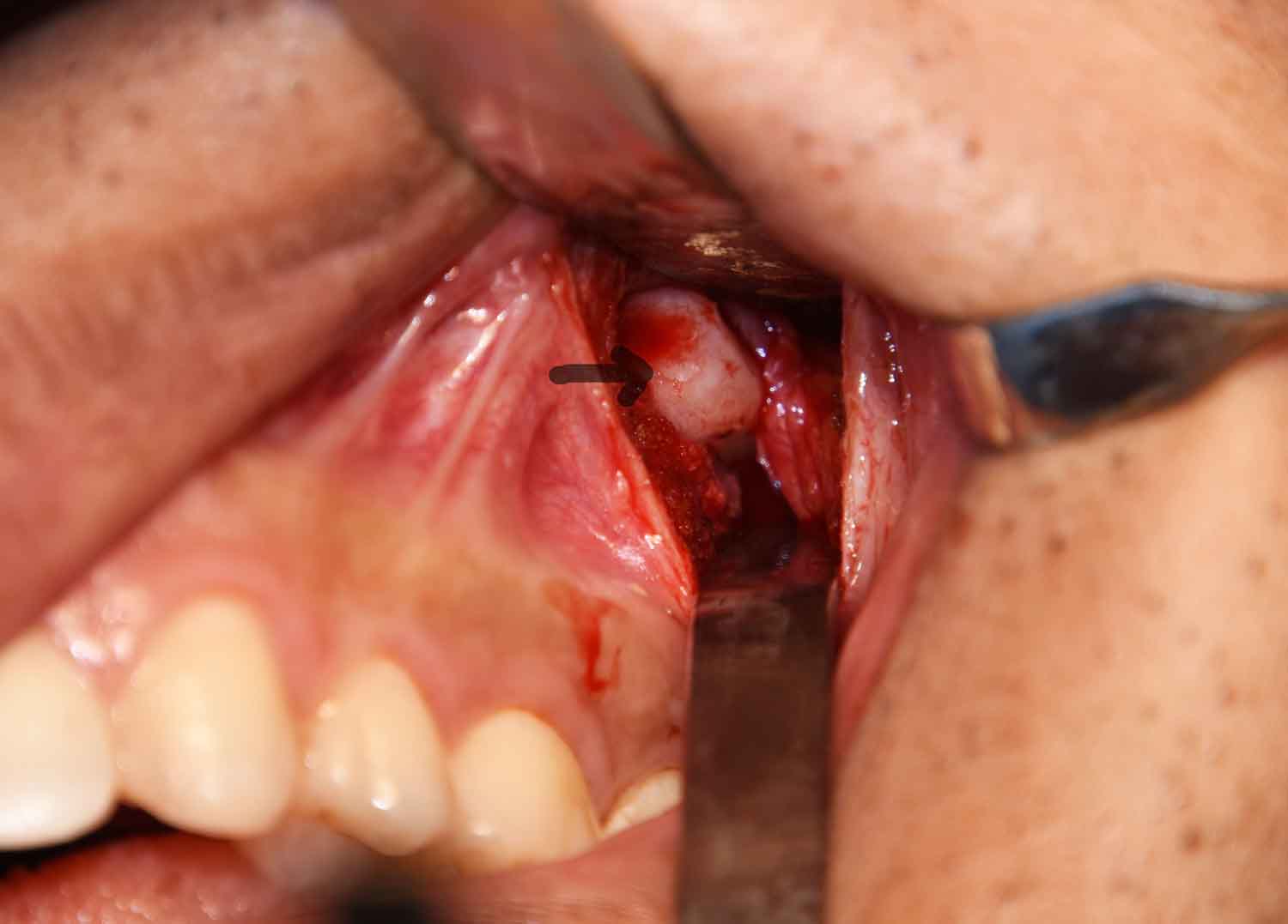
Correction of this problematic full midface fullness can be done by an intraoral posterior maxillary osteotomy and infracture. Through an intraoral approach and a small vestibular incision over the palpable bony bump, the base of the posterior maxillary protrusion is fractured back into the maxillary sinus. This changes the unnatural bony protrusion back into a more natural concavity and eliminates the external lower cheek fullness.
Dr. Barry Eppley
Indianapolis, Indiana