

 There are numerous types of congenital ear deformities. The constricted ear represents a tightness of the ear like a cinch around the outer helix of the ear which makes it smaller and often folded somewhat onto itself. The Stahl’s ear deformity, also known as a Vulcan or Spock ear, has a cartilage fold that can cause a pointed malformation in the upper part of the ear. Both types of ear deformities are uncommon but are even more rare when they occur together.
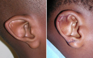
In the July 2015 issue of the International Journal of Plastic Reconstructive and Aesthetic Surgery, an article appeared in print entitled ‘ Surgical Correction of Constricted Ear combined with Stahl’s Ear’. Over a seven year period, the authors had 19 patients with constricted ear with Stahl’s ear, most of whom had it on just one side. They were surgically treated by a technique that consisted of an initial double Z-shaped skin incision made on the back side of the ear with the entire layer of cartilage cut parallel to the helix traversing the third crus to form a fan-shaped cartilage flap. The superior crus of the antihelix were shaped by folding the cartilage rim. The cartilage of the abnormal third crus was made part of the new superior crus of the antihelix and the third crus was eliminated.
Postoperative assessment of the ear reconstructions based on symmetry, helical stretch, successful elimination of the third crus, the auriculo-cephalic angle, and the substructure of the reshaped ears. All reconstructions were rated as excellent to good without any complications seen. This study shows that even the rarest of congenital ear deformities can be successfully treated with the proper surgical technique.
In the human ear the bifurcated Y-shaped superior and inferior crus is a major component of its upper half. In Stahl’s ear deformity an aberrant crus usually replaces the superior crus, crossing the scaphoid fossa from the site of the normal bifurcation of the antihelix posteriorly towards the helix which gives it an abnormal J-shape. A surgical technique to correct Stahl’s ear deformity can be done by a Z-plasty to the incision to lengthen the skin on the posterior surface, together with making use of the aberrant crus to reconstruct the superior crus without reducing the ear size using horizontal mattress sutures. This is achieved by posterior scoring and suturing without any cartilage excisions which converts the J antihelix into a Y antihelix.
Adding the constricted ear problem to the Stahl’s ear raises the stakes in terms of reconstructive difficulty. The tightness of the skin and shortage of cartilage necessitates the need to release the constricted cartilage into a fan shape and use Z patterned incisions on the back of the ear.
Dr. Barry Eppley
Indianapolis, Indiana