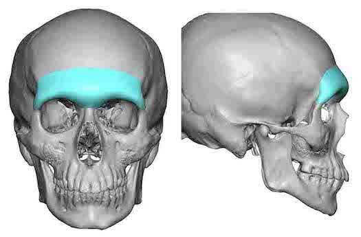
One distinct male feature is the of a prominent brow bone at the lower end of the forehead. It is one of many facial features that distinguishes males from females. It extends over the eyes with a horizontal projection, has a brow bone break before heading up into the forehead and has a slight glabellar depression between the medial brow bone prominences.
In today’s male beauty trends many models display a very strong or exaggerated brow bone appearance. It borders on being an almost angry appearance but is better anatomically described as an ultra low brow bone projection with near complete coverage of the upper eyelids. While I frequently get requests for such a brow bone augmentative change, I have to advise that such requests are almost always impossible to achieve. This is due to the fact the soft tissues of the eyebrows are tight and can not be driven down below the existing brow bones unless these tissues have been first expanded before a brow bone implant is placed.
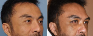
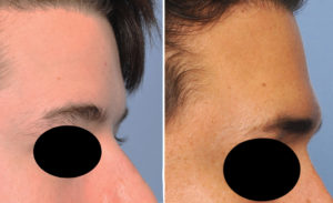
More realistic brow bone augmentation results create less dramatic changes but appreciable improvements nonetheless. More horizontally oriented projection across the orbital rims, a brow bone break and a more distinct corner or tail of the brow bone are achievable augmentative changes.
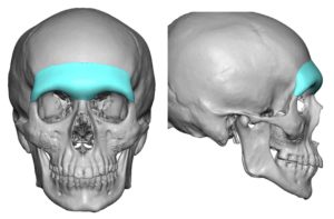
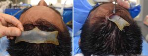
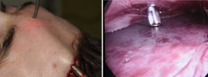
Custom brow bone implants usually require a three incision placement technique, all of which are small and heal inconspicuously. The key is to get the implant low enough over the brow bones through adequate soft tissue release and good positioning and fixation over the lateral orbital rims. One may think this can be done exclusively from a superior approach only…but that can be ill-advised if the brow bone implant goes any length along the lateral orbital rims.
Dr. Barry Eppley
Indianapolis, Indiana

