Background: The shape of the head is largely controlled by the contour and thickness of the various skull bones. The lone exception is the side of the head from the lateral orbital rim all the way to the back of the head on which the temporalis muscle runs. It is the thickness of this muscle combined with that of the underlying temporal bone that creates the shape of the size of the head.
The temporal muscle is a broad fan-shaped structure that has its origin on the temporal lines of the parietal bone and inserts to the coronoid process and retromolar fossa of the mandible. Its action is to elevate and retract the lower jaw. But the muscle exhibits tremendous differences in thickness, being very thick by the side of the eye due to the concavity of the temporal fossa and becoming thinner as it crosses the convex shape of the temporal bone above the ear. Despite it being thinner above the ear it is still upwards to 1 cm in thickness in males. This makes a major contribution to the shape of the side of the head at the vertical ear level.
In men due to shorter hairstyles the shape of the side of the head is very visible. Its contour and relationship to the projection of the ear is evident from the frontal view. When it is excessively wide, which is usually seen as a prominent convexity, it may become aesthetically bothersome. While many think that temporal bone reduction is the solution to making the side of the head straight (non-convex), it is the muscle that is more easily removed and effective without any adverse function effects on lower jaw movement.
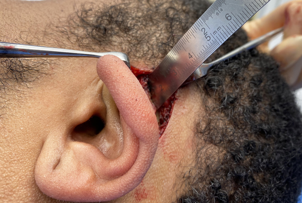
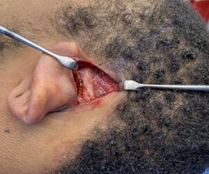
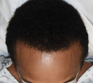
Case Study: This male was bothered by a wide side of the head which was overly convex. The definitive test to demonstrate the contribution of the temporal muscle is to widely one’s mouth and see how that affects the side of the head shape. If it becomes thinner/more straight then one knows muscle removal in that area will produce an even greater thinning effect. He had a positive temporal test.
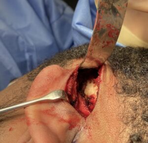
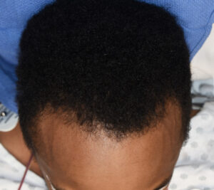
One of the common questions about this type of temporal reduction surgery is whether there are any adverse effects on lower jaw function. And if not how can that be since surely the muscle is there for a reason. To understand why there is no loss of function it is important to look at the anatomy of the temporal muscle. The temporal muscle can be divided into two parts, anterior and posterior. The very large and thick anterior portion of the muscle (about 70% of its muscle mass) runs vertically and is primarily responsible for bringing the lower jaw back into occlusion. (elevation of the mandible) The posterior portion of the muscle runs horizontally of which its contraction results in recursion of the lower jaw. There is actually an intermediate or middle portion of the muscle whose muscle fibers run obliquely whose contraction creates both elevation and retrusion of the lower jaw as well as lateral movement. While the posterior portion of the muscle covers a large surface area its total contribution to the overall muscle mass is less than 20%. As a result its loss does not create any visible effects on widely opening and closing of the lower jaw.
Case Highlights:
1) A wide or convex side of the head is seen in the frontal view and has a major contribution from the temporal muscle that runs across the convex temporal bone below it.
2) The postauricular incision provides direct access for temporal muscle removal and does so in a scarless manner.
3) The typical thickness of the posterior portion of the temporal muscle above the ear can be as much as 10mm or more in some patients.
Dr. Barry Eppley
Indianapolis, Indiana

