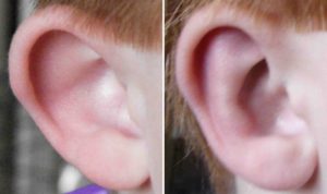
Otoplasty, often referred to as ear pinning, remains as one of the most satisfying of all plastic surgery procedures above the shoulders. In a very short operative time, the ears can be dramatically reshaped to assume a less noticeable and more aesthetically pleasing appearance. Through an incision on the back of the ear, the otoplasty procedure is performed without visible scars.

Some cases of protruding ears are the result of a large concha or bowl, not the antihelix. The inner tier, or third level, of the ear is shaped like a bowl and helps capture sound to direct it into the ear canal to the ear drum. The concha forms the under support for the outer antilhelix and helix. When it is too large, it can be the primary source of a cosmetic deformity. When this is present, one will often have a good antihelical fold but the ear still sticks out too far. Without reduction of the large concha, other suturing methods will be unsuccessful. Removing a wedge of conchal cartilage and using sutures that pull back the concha towards the mastoid are needed to make the ear sit closer to the side of the head.
Many otoplasty procedures require a combination of antihelical and conchal manipulations to create a new ear position that does not look deformed or ‘crimped’. I have seen several cases of ears pulled back too far that had unusual or unnatural folds in them. This is the result of not identifying the total cartilage problem and trying to make one cartilage reshaping method (usually antihelical fold suturing) do too much. As a general rule, the helical rim should always stick out just beyond the antihelical trim in a frontal view. And there should always be a small vertical curve or slope from the antihelical rim to the base of the concha without a sharp transition or indent in the skin. Such deformities can be seen intraoperatively and one should not expect much relaxation of the ear shape after surgery. In short, don’t overcorrect counting on it ‘evening out’ after surgery.
The complex cartilage shapes of the ear usually defy a simple suture or two to adequately reshape them. Appreciating what cartilage abnormalities makes the ears stick out too much will enable the right combination of cartilage bending and resection to give the ear a better profile without deforming it. The repositioning of the cartilage through sutures allows scar tissue to form which is ultimately responsible for the long-term retention of their altered shape.
Dr. Barry Eppley
Indianapolis, Indiana


