

Background: Osteomas are benign bony growths that develop on the outer surface of a bone. (outgrowth of the outer cortical layer) While they can occur anywhere on the body they are very common on the skull with the forehead being a frequent location. Their cause is always clear and many occur for no obvious reason. Trauma is frequently cited as a cause, and in some patients that event can be traced to the osteoma, but the vast majority of them have no obvious reasons for their presence.
The frontal skull osteoma is the most common type that presents for removal due to its easy visibility on the exposed forehead. incision over it provides the most direct method of removal with full visibility of it. And while this may be acceptable for some patients if they have a horizontal wrinkle line near it most patents would prefer to avoid any sign of a visible scar. Thus an endoscopic technique is preferred which requires to technical expertise to perform.
One unique feature of many osteomas is their pedunculated feature. This provides a convenient place for an osteotome to separate it from the underlying skull where it can be grasped and removed. But that alone does not provide assurance of a complete removal with a flat surface or eliminates the risk of recurrence. Rasping the removed osteoma base and sealing it with bone wax provides the final smoothing effect.
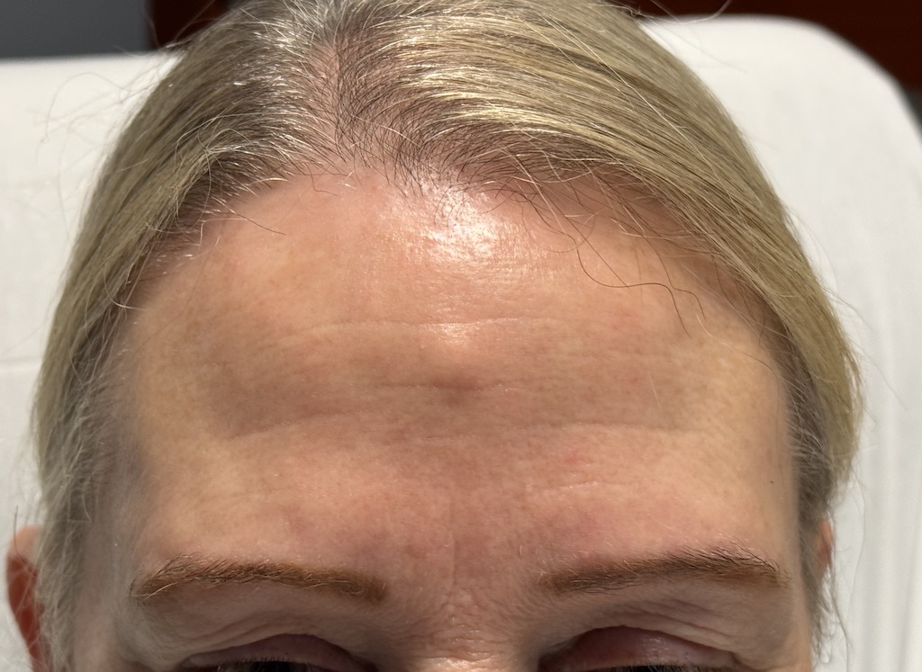
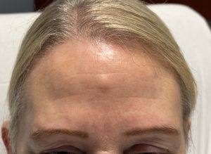 Case Study: This female developed a dead center of the forehead small osteoma over a five year period. There was not history of trauma and she was unsure if it was still growing or not. While she did have some horizontal wrinkle lines that could be used for its removal, she preferred to have an unscarred forehead.
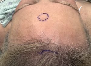
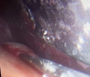
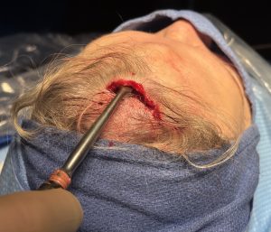
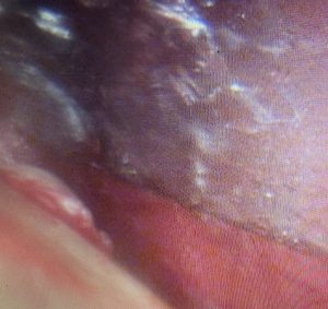 Under anesthesia a small scalp incision was made right behind the frontal hairline. Using a 30 degree endoscope the central forehead osteoma was visualized.
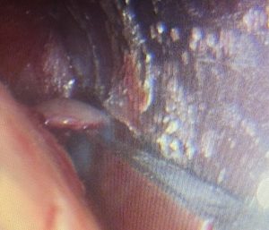 Using an osteotome (chisel) the bony mass was removed from the underlying skull surface. This was easily done since then osteoma had a pedunculate shape. It was then grasped and brought out through the incision.
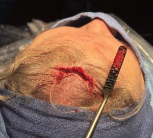 To remove any remaining osteoma a #8 rasp was used to smooth down the bone surface down. A small amount of bone wax was compressed over the bone removal site to prevent any chance of regrowth.
 The scalp incision was closed with small dissolveable sutures and a forehead wrap applied.
The forehead osteoma may be a benign outcropping of bone growth and its appearance can make it look like a ‘third eye’ sometimes but many patients do not want a visible scar tradeoff for its removal. The endoscopic technique provides a method for avoiding that adverse aesthetic tradeoff.
Key Points
1) Osteomas of the skull are not uncommon with those occurring on the forehead as the most aesthetically concerning.
2) While an incision over the osteoma is the most direct approach patients usually prefer a remote incision endoscopic scalp approach.
3) An osteotome and skull rasp are needed through the remote scalp incision for a complete smoothing effect.
Dr. Barry Eppley
World-Renowned Plastic Surgeon