


Background: Of the five surfaces of the skull the sides of the head make up two of them and are the only surfaces covered by muscle. They are also the only skull surfaces whose profile (shape) can only seen from the front and back views of the head. As a result abnormal shapes to the side of the head are usually seen as either too wide or too narrow. (besides asymmetry) While too narrow of a head shape is usually due to a lack of bone projection/development the too wide head shape can be the result of excessive temporal muscle thickness.
Such muscle thickness can be reduced to make the head less wide which is the basis of the temporal reduction procedure. By removing the full thickness of the posterior portion of the muscle and reducing the thickness of the larger anterior muscle portion the head shape can be narrowed and /or its convex shape reduced. The temporal muscles can be so manipulated without compromising jaw function…which is usually the most common patient concern about the surgery.
In some wide head patients the ears may also be pushed outward or have an increased auriculocephalic angle. This is presumably not a coincidence and is due to developmental proximity. Even in borderline upper ear protrusions the concern with temporal reductions is as the side of the head shape becomes more narrow the natural position of the ear will then look more protrusive. Thus an otoplasty procedure can be considered along with the temporal reduction done concurrently. Then the only question is should the otoplasty be done through the postauricular incision used for the temporal reduction or should a separate ear incision be made.
Case Study: This patient had a convex head shape that is often referred to as light bulb shape. The convexity is higher up on the side of the head closed to the bony temporal line than the ear. The upper ears were also slightly protrusive. The decision was to set the upper ears back as they were already protrusive even before the side of the head was narrowed.
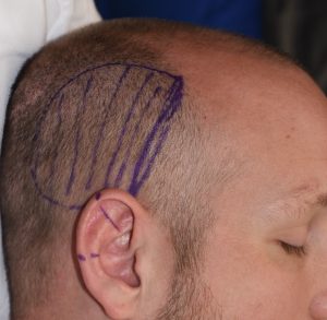
 Under general anesthesia the zone of temporal muscle removal was marked in the side of the head and the markings on the ear were for the upper and lower limited of the ear protrusion.
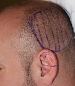
 Through postauricular incisions the posterior temporal muscles portions were removed. When seen from above the top of the ears were more protrusive than before.
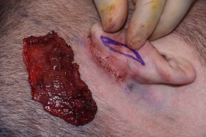
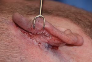 Then through a second incision higher up on the back of the ear sutures were used to set the upper ear back.
All incisions were closed with dissolvable sutures and a drain placed for the temporal reductions which was removed the following days.
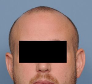
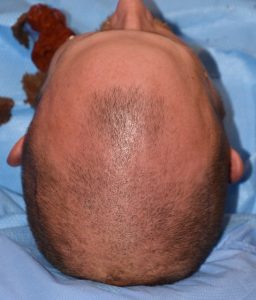
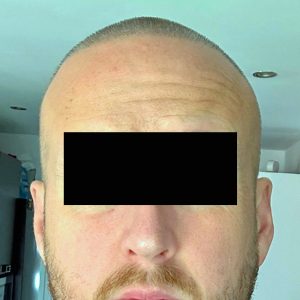 Long term followup supplied by the patient over six years later showed a less wide head shape and ears that did not stick out.
Developmentally it is no surprise that in some patience with a wider Side of the head the underlying ears may also stick out as well. Even when the ears are marginally protrusive a significant temporal reduction can make them appear even more so. Thus there is always a consideration for reshaping the ears when a side of the head reduction is done. In most cases this is not necessary but if the patient has a preoperative concern about whether the ears will stick out further after the procedure then they undoubtably will.
In performing combined setback otoplasties with temporal reduction the primary consideration is incisional placement. There is already an incision in the postauricular sulcus and it is tempting to think that ear cartilage manipulation can be done using it. This may be true if the primary mechanism of the ear setback is a conchal repositioning by suturing it to the mastoid fascia. But when the primary method to successfully set back the ears is closer to the helical rim a more direct incisional approach is going to be needed. While these two incisions are relatively close the superb vascularity of the ear permits that proximity do not be a problem as long as the incision up on the back of the ear is not too long and the amount of skin undermining is not extensive.
Key Points:
1) For the convex side of the head shape subtotal removal of the temporal muscle produces a less wide and more straight appearance.
2) A wide side of the head can be associated by developmental proximity with some upper ear protrusion.
3) Temporal reduction and setback otoplasty can be performed at the same time through separate incisions.
Dr. Barry Eppley
World-Renowned Plastic Surgeon