

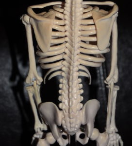
Rib removal surgery for horizontal waistline reduction involves modification of the lower three bony ribs. (#s 10,11 and 12) While the bottom two ribs are the free floating ribs (as they have no distal attachments), rib #10 is an attached rib. Its bony portion wraps around the torso to become cartilage where it continues to blend into the subcostal collection of ribs which attaches to the sternum.
While these anatomic differences between ribs #10 and 11 and 12 seem merely descriptive in nature, they have significant surgical relevance in rib removal surgery. In looking at many different anatomic illustrations I have always been impressed that what is illustrated is often not what I see in surgery.Typical illustrations have these ribs as being parallel to each other and the rib lengths of 11 and 12 being nearly equal. Yet in surgery I have always been impressed with the significant downward angulation of ribs 11 and 12 (compared to rib #10) and how short rib #12 is compared to #11.
 Then the other day my assistant brought in a small anatomical model she found in a garage sale. There were many proportions in the model that were distorted but the lower ribcage showed perfectly what I always see in surgery.
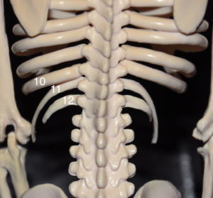
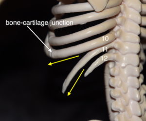 Both ribs #11 and #12 were significantly angulated downward with rib #10 being much more horizontal. This makes sense since without the need to wrap around the torso to become part of the subcostal ribcage there is no benefit to being more horizontal in orientation. Rib #12 was also shown as being much shorter than #11. This is always seen is surgery where the length of rib #11 removed is at least 5 to 6 cms longer than that of #12. Rib #10 wraps around and becomes cartilage at the costochondral junction (bone-cartilage junction) and is where the rib is disarticulated distally during the surgery.
It is surprising to see an inexpensive anatomical model, found in a garage sale, that actually reflects what I have seen over and over in rib removal surgery. And shows what I usually do not seen in many established illustrations of the lower ribcage.
Dr. Barry Eppley
Indianapolis, Indiana