

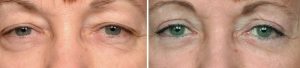
Blepharoplasty, or eyelid tucks, remain the mainstay of rejuvenation of the aging eye area. Loose eyelid skin, herniated fat, and wrinkles are all largely tissue issues that respond best to surgical manipulation. Adjunctive techniques such as browlifts, ptosis correction, chemical and laser peels are all options that may complement the effects from a blepharoplasty either through aesthetic enhancement or improvement of pre-existing asymmetry.
One of the most common complaints after eyelid surgery is asymmetry. Because the eyelids drape over the eyes, it is most visually analyzed area of the face…particularly after having spent money and gone through swelling and bruising to get an improved appearance. This critical analysis is exacerbated as many women use magnifying mirrors for applying makeup. Concerns usually involve issues of upper eyelid incision placement, residual skin and fat pockets on both the upper and lower eyelids and differences in lid position. In my experience, it is the upper blepharoplasty that generates the most asymmetric concerns after surgery.
 The best way to avoid postoperative asymmetry concerns of the eyelids is to recognize that it exists beforehand. Almost everyone has some degree of eyelid asymmetry and it is important to carefully look for it during a consultation. Differences often exist in eyebrow position, amount of redundant upper eyelid skin and lid position on the globe. Pointing it out to patients with a mirror and a q-tip usually demonstrates a novel finding to them. Almost everyone has some degree of upper eyelid asymmetry just as they have other areas of facial asymmetry.
One of the main causes of upper eyelid asymmetry is differences in the distance between the eyebrow and the lid margin. This can be caused by eyebrow asymmetry, lid margin asymmetry (ptosis) or differing amounts of redundant upper eyelid skin weighing down on the eyelid. Eyebrow asymmetry, if significant, can be managed by a one-sided endoscopic browlift technique. However an endoscopic browlift is not a prevision procedure that lends itself to predictable changes at the millimeter level. So its use should be carefully applied as it is easy to create brow asymmetry in the opposite direction. Small amounts of lid sag can be managed by microptosis correction done through a posterior or internal approach. (e.g., Fansenella-Servat technique) Asymmetric eyelid skin removal is achieved by careful preoperative measurement using calipers and loupe magnification.
One major avoidable cause of upper blepharoplasty asymmetry is incision placement. Having incisions at two different levels on the eyelid is unavoidably noticeable. Carefully looking for the natural upper eyelid skin crease and assuring symmetry through caliper measurements prior to surgery is important. Asymmetric eyelid incision placement is very difficult to correct afterwards so it is best prevented.
 Another cause of asymmetry and concern is redundant skin or a dog ear at the medial aspect of the upper eyelid incision. The extent of the inner aspect of the upper eyelid incision is controlled by proximity to the nose. There is a point where the incision should not cross onto the nasal skin. But redundant upper eyelid skin often hangs near the nose. This can leave a bag or roll of skin at the end of the incision if it is not carefully looked for and worked out during the procedure. This can also be corrected secondarily as well.
Lower blepharoplasty asymmetry is usually due to two issues, lower lid position and residual prominent fat pockets. Lower lid malposition or ectropion is the most common lower blepharoplasty problem and one that can cause eye symptoms as well. (irritation, tearing) Its prevention and secondary treatment have been well described through lid tightening, suspension and canthal manipulation techniques. Residual, missed or asymmetric fat pocket removal can become apparent months after surgery when all the swelling has subsided. They usually occur in the medial or lateral fat pocket areas and can be corrected secondarily through transconjunctival removal or cautery ablation.
Dr. Barry Eppley
Indianapolis, Indiana