

Background: The angle of the jaw is a unique facial bone area as its shape is highly influenced by the natural development of the jaw and the masseter-pterygoid muscle sling which is attached to it. It is the most recognized are of the ramus portion of the mandible.Regardless of the actual angle shape, it always has a posterior and inferior border which relate to each other through an angular relationship.
The sagittal split osteotomy (SSRO, sagittal split ramus osteotomy) is the most common orthognathic surgery technique of the lower jaw. It is a very clever and technically sensitive technique in which the the ramus of the mandible is cut longitudinally, splitting it into an outer proximal segment and an inner distal segment. This bone sectioning allows the tooth-bearing portion of the lower jaw (distal segment) to be moved forward or back depending upon the desired occlusal positioning. Its main benefit is that it allows maximal bone contact of the moved bone segments for rapid healing and bone stability.
While in theory the shape of the jaw angle is not changed from an SSRO procedure, this is not always true. The shape of the angle can end up altered due to a ‘bad split’, devascularization of the bone segment and how the proximal and distal segments are put back together. This risk escalates when a second SSRO procedure for revisional orthognathic surgery due to a malocclusion or undesired facial skeletal changes.
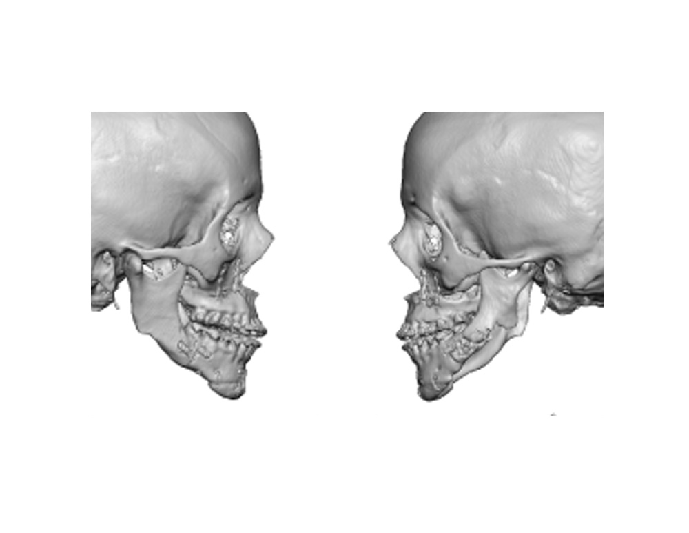 Case Study: This 35 year-old female had a history of two prior orthognathic surgeries, both of which included SSRO procedures. She was bothered by the lack of jaw angles, a sunken facial contour near the ears and facial asymmetry. The loss of jaw angles was greater on the left side of her face with the additional loss of the entire lower border of the ramus as seen on a 3D CT scan.
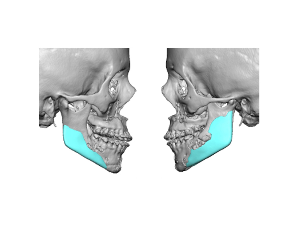
 The 3D CT scan was used to create custom jaw angle implant designs which recreated a defined jaw angle shape as well as symmetric angles and ramus bone segments. These designs were used to make actual implants made out of medium hard durometer silicone.
Under general anesthesia, she had her custom jaw angle implants placed through her original intraoral posterior vestibular incisional scars. The scar tissue was quite adherent and her fixation plates were visualized and maintained. Once positioned the implants were secured with multiple 1.5mm titanium screws placed percutaneously.
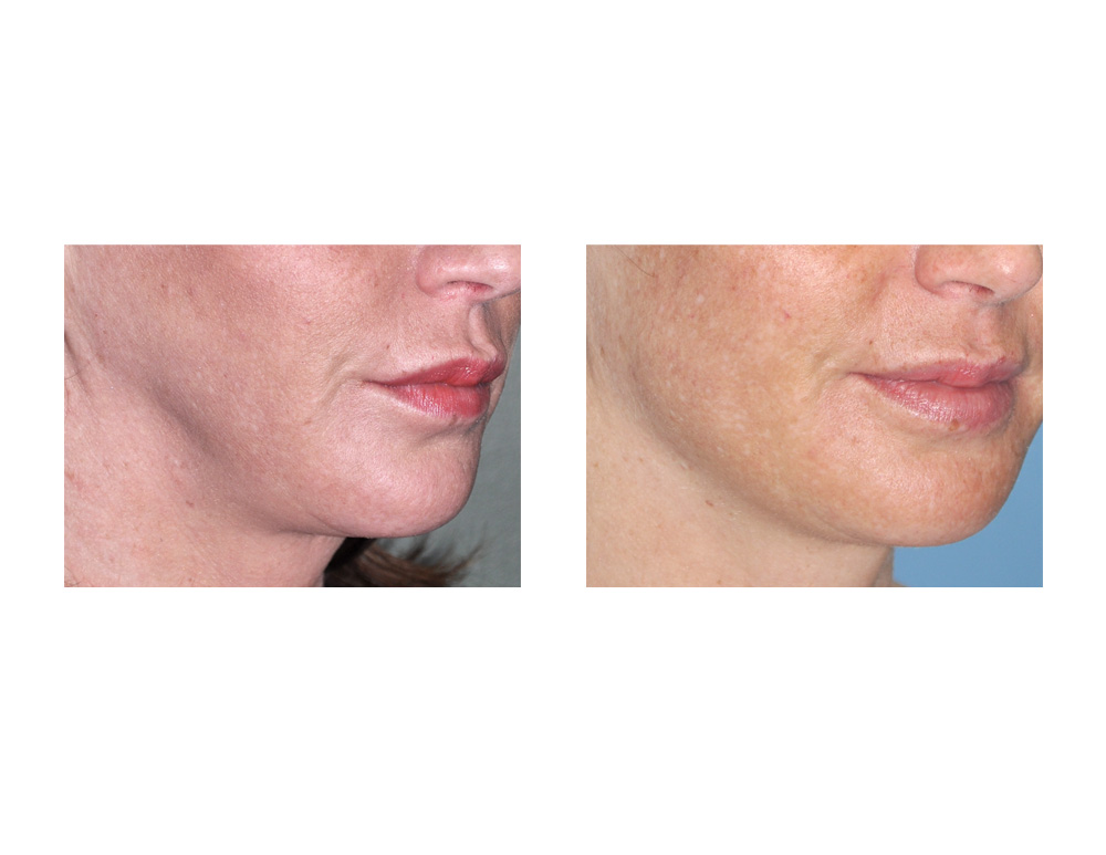
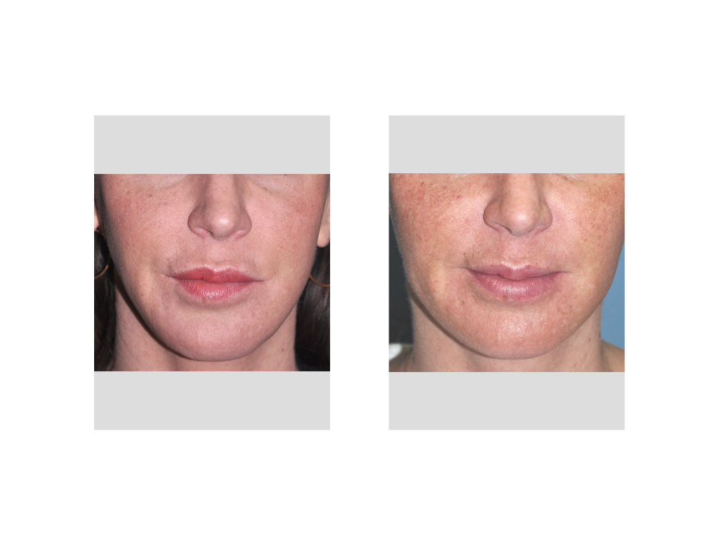 After considerable facial swelling resolved, her restored jaw angles/mandibular rami provided a much improved lower facial contour and fullness. Sharp well defined jaw angles were not created as this was not what her original face looked like. She just wanted normal fullness over the jaw angles that was a lot more symmetric.
Case Highlights:
1) The shape of the jaw angle and the posterior inferior border of the mandible can be altered by sagittal split osteotomies for orthognathic surgery.
2) Reconstruction of jaw angle deformities is best done by having custom jaw angle implants made from the patient’s 3D CT scan.
3) When designing custom implants for surgically created jaw angle defects it is important to account for fixation hardware and what the shape of the original jaw angle may have been
Dr. Barry Eppley
Indianapolis, Indiana