

Background: Osteomas are well known benign bony growths which can occur from within, on or even outside of the bone in the surrounding soft tissues. In the craniofacial skeleton, osteomas usually occur as a peripheral lesion arising from the periosteum. While there are multiple etiologies for them the most recognized is that of trauma. The mechanism is that a subperiosteal bleed occurs which serves as the stimulus for bone formation. This is not infrequently seen on the forehead and skull.
While the rest of the face is certainly exposed to trauma the occurrence of osteomas is far more rare. One of the rarest facial locations is on the zygoma, specifically that of the zygomatic arch. It is perhaps the very thin bone of the arch and its very limited surface area that limits its potential for a subperiosteal bleed and osteoma formation.
The presence of an osteoma is detected by an external contour change. It is a very slow growing bony lesion which will feel very hard and immobile. While removing such osteomas as straightforward the challenge is often getting there to do it.
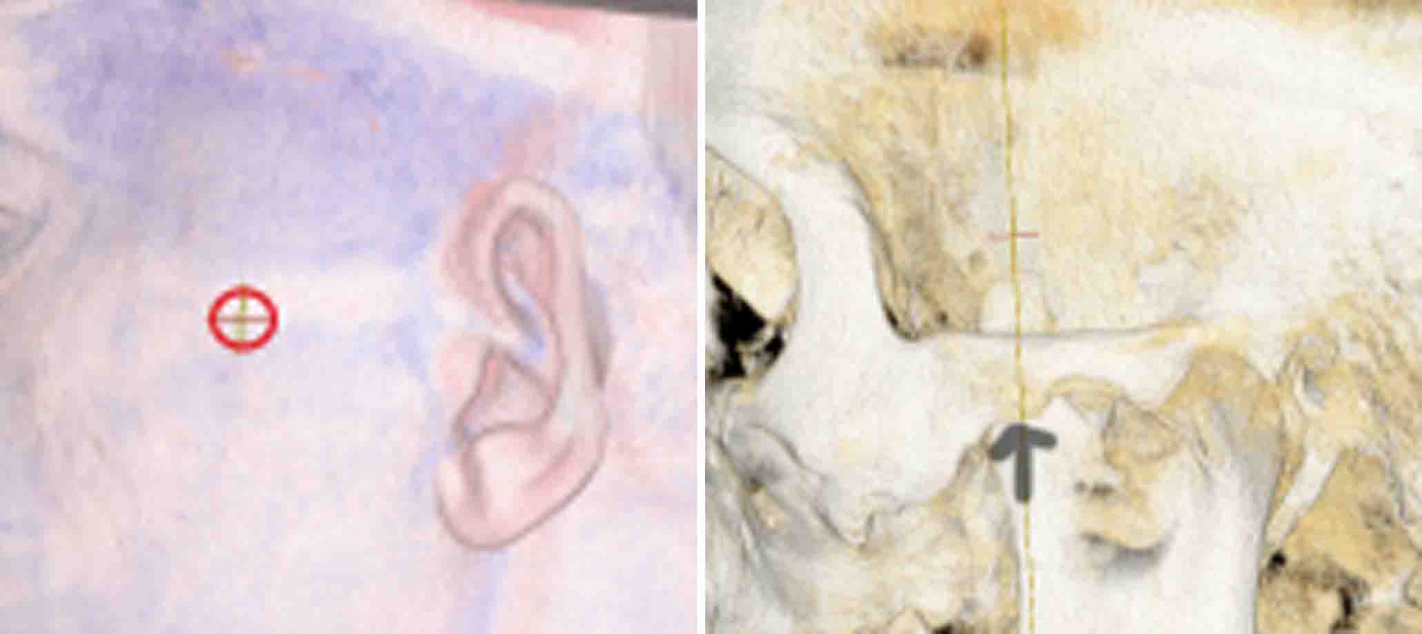 Case Study: This young male sustained blunt trauma to the left side of his face over the cheek/zygomatic arch area. When all the swelling finally resolved he was left with greater fullness over the area which was asymmetric to the opposite side of his face. It felt firm and stuck out more. A 3D CT scan showed a concentric bony mass on the inferolateral side of the zygomatic arch right at the suture line.
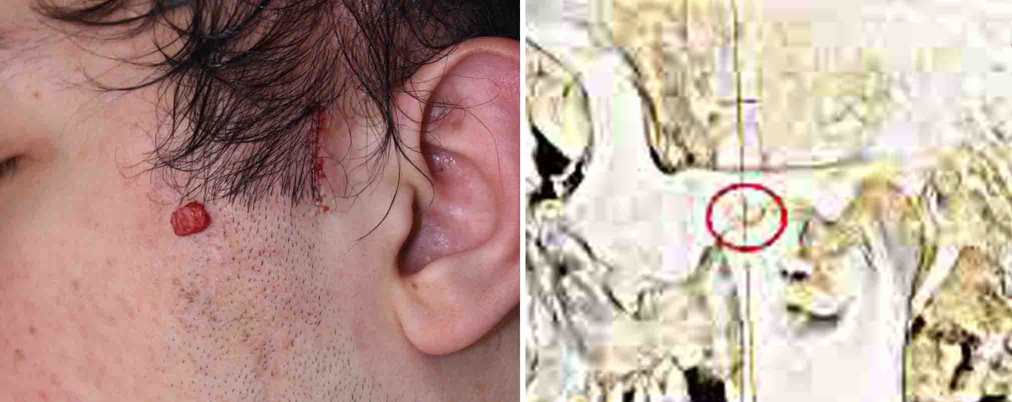 Under general anesthesia and through a posterior sideburn hair incision the temporal process of the zygomatic arch was initially exposed. Moving forward along the arch the bony bump was identified and removed. A posterior arch osteotomy was also done and infractured 4mms to ensure that the arch asymmetry was improved.
It is likely in this case that the trauma caused a subperiosteal bleed at the mid-arch suture which may have become slightly disrupted. This was enough to cause a small osteoma to form which contributed to the persistent facial fullness/asymmetry. Access to the middle portion of the zygomatic arch is difficult through any approach other than a coronal scalp incision. But such an approach would be excessive given the relatively modest size of the osteoma.
Case Highlights:
1) Trauma and subperiosteal bleeding is a well known stimulus for the development of an osteoma on the face.
2) Traumatic zygomatic arch osteomas are very rare and can occur at the mid-arch suture.
3) Removal of zygomatic arch osteomasis best done through a posterior sideburn hair incision.
Dr. Barry Eppley
Indianapolis, Indiana