

Background: The finding of a bony growth on the palate is most commonly the result of the development of a tori. They typically present as a small midline bony lesion less than a few centimeters in size in early adult life. They are capable of growth over one’s lifetime but it its usually very slow.
Palatal tori are very common and are estimated to occur in 10% to 20% of the general population. They are more prevalent in some ethnic populations but are also more often seen in females. It is not known why they occur but they can be inherited and have been claimed to be an autosomal dominant trait.
Palatal tori occur in different patterns and are classified based on their shape. They can occur as flat, spindle, nodular and lobular types. Nodular and lobular tori are usually bigger in size. Palatal tori are benign bony outcroppings that do not usually need removal unless they are a source of irritation or are an obstruction to the fit of dentures.
Case Study: This middle-aged female had large palatal for her entire life. She had a family history of her mother having large palatal tori as well. They occasionally became sore but she never considered removing them until she needed partial dentures. Her palatal tori consisted of four bony growths, two large smooth ones on the inside of the alveolar ridge at the molar area as well as a midline lobular one that had two large halves. All four tori merged together leaving just a narrow linear gap between them. (a source of food entrapment)
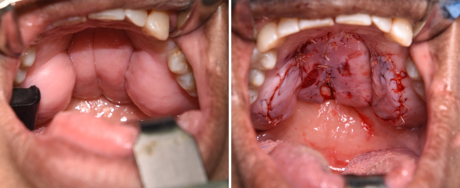 Under general anesthesia, laterally based mucoperiosteal flaps were used to expose the lateral tori for their reduction by a high speed burr and osteotomes. A t-shaped mucoperiosteal flap was used to exposed the bilobed midline tori for their reduction by a similar bore removal technique..
Massive palatal tori removal requires most of the hard palatal tissues to be elevated for exposure and adequate bone removal. While effective, complete mucosal healing requires a few weeks and is fairly sore.
Highlights:
- Palatal tori are commonly a single midline bony lesion but four in a single patient is very rare.
- Removal of palatal tori may be necessary for the fabrication of partial or full dentures.
- The removal of palatal tori requires the reflection of full-thickness mucoperiosteal flaps.