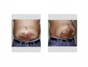
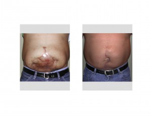
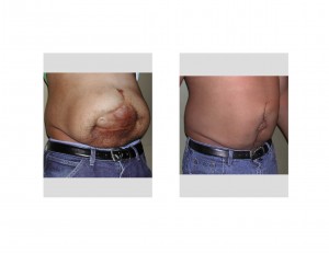
Background: Abdominal wall disruptions and deformities occur from a wide variety of problems. But infection from traumatic injury, surgery or a bowel-related disease process can produce a devastating and life-threatening abdominal wound problem due to having to be packed open. Once the infection is cleared, the open abdominal wound can usually be secondarily closed once tissue inflammation has subsided but, in some cases, the open wound is better to be skin-grafted after developing a good bed of granulation tissue. This is an older school of surgical thought but a very safe one once the abdominal wall or intraperitoneal cavity has been infected.
A large abdominal wall deformity with a wide scar and underlying separation of the rectus muscles poses a daunting task for achieving a more normal looking abdominal wall. While the underlying hernia, or the potential for it, is difficult enough the scar width seems almost impossible that it can ever be significantly narrowed.
The key to repair of large secondary abdominal wall problems lies in the rectus muscles. Any hope of simultaneously fixing the hernia and narrowing the scar lies in their approximation. The muscles must be adequately mobilized and brought together in the midline, for by so doing it carries with it he overlying skin. Ideally, patients should be six months or more after their initial event to allow the surrounding tissues to become more lax. In some patients, weight loss can also be helpful to reduce the underlying pressure on the closure. (although many patients have actually gained weight from their pre-injury state due to less mobility and an inability to exercise)


Case Highlights:
1) Large abdominal wounds that have been allowed to heal and skin grafted are associated with underlying hernias and can appear impossible to ever regain a more normal looking abdominal wall.
2) Large abdominal scars with a hernia can be successfully repaired with scar excision, rectus muscle reapproximation and abdominal skin flap mobilization.
3) Such midline tissue tension on the abdominal wall can be expected to result in some scar widening and surrounding abdominal tissue distortions that can benefit from a secondary scar revision.
Dr. Barry Eppley
Indianapolis, Indiana


