Background: Prominent brow bones are the direct result of the development of the underlying frontal sinus. While all of the frontal forehead bone above the brows is very solid and thick skull bone, the brows are made up of air with only thin bone in front and back of its thickness. The anterior or frontal part of the brow bone beneath the eyebrows is remarkably thin, often only being a few millimeters thick.
Brow bone reduction is done for two main reasons. Men who have large and very prominent brow bones often want them reduced to look less ‘Neanderthal-like’. Women with larger brow bones or men to women transgender patients who want a softer and more feminine appearance may want their brows reduced and the tail of the brow bone reduced and flared upward. In some cases simple burring may be effective to achieve these goals but most of the time the outer table of the frontal sinus bone must be removed and reshaped to get a significant reduction. The thin outer bone of brow bone makes only a few millimeters reduction possible with burring.
When the frontal sinus is enlarged, it most always involves both sides of the brow bones. This is because the frontal sinus in most people is paired and exists under both eyebrows. But the frontal sinuses are rarely symmetrical and the septum that exists between them frequently deviates to one or other side, allowing for one frontal sinus to become larger than the other. This can account for the rare occurrence of asymmetrical brow bone hypertrophy.
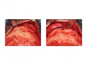
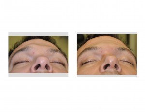
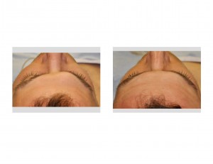
Case Study: This 33 year-old male had one enlarged brow bone that had bothered him for years. He had no specific history of trauma to the area. It had just developed naturally that way. It created the appearance of a large knot or ball on his brow that also pushed down into the eye socket, giving it a swollen appearance. He had no pain or numbness over the brow area.


Case Highlights:
1) Brow bone hypertrophy most commonly occurs on both sides and rarely on just one side.
2) Brow bone reduction is done through an open coronal (scalp) approach by removal and reshaping of the bone overlying the enlarged frontal sinus.
3) Brow bone reduction has no adverse effect on the frontal sinus.
Dr. Barry Eppley
Indianapolis, Indiana


