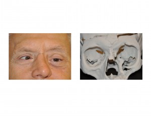
Background: Orbital defects are common from a variety of causes. Whether it be trauma (orbital floor fractures), congenital defects or tumor resection, the most common problem is that of loss of bone support at the floor level. As the bony orbit is a box (really a pyramid turned on its side), lowering the floor affects the position of its primary content, the eye. As the eye drops down, it causes double vision as the eyes no longer have binocular visual capability.
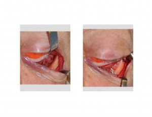
Reconstruction of the orbit floor has been successfully done by about every conceivable method. From autogenous bone grafts to synthetic implants, these ‘spacers’ build up the orbital floor and bring the eye up to a position closer to a horizontal level to the opposite eye. While there are advocates for each orbital reconstruction method, the frank answer is the best method is the one that works. The surgical method is secondary to the skill and experience of whomever performs it.
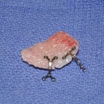
One craniofacial reconstruction method not commonly used or discussed, but equally effective, is that of fabricating a computer-generated implant. Using a 3-D model made from a patient CT scan, an implant can be made that mirrors the bone level of the opposite side. While this approach takes more time and expense, the exactness of this method may be justified in more complex orbital deformities.


There was a fair amount of orbital swelling as expected that required about six weeks until the tissues returned to normal. At that time, the eye was at a completely horizontal level to the opposite eye. The double vision was not completely eliminated but it was much improved.
Case Highlights:
1) Orbital reconstruction, particularly that of the lose floor bone, can be successfully done by a variety of materials. Getting the floor back up to a level to the opposite side can help improve double vision symptoms.
2) Custom HTR implants can be made from a 3-D model created off of the patient’s CT scan. While some adjustment of the implant is almost always necessary, one has a good place to start that is anatomically accurate.
3) The end goal of orbital floor reconstruction is even eye levels between the normal and deformed sides. Success is defined by that criteria, not the method used to get there.
Dr. Barry Eppley
Indianapolis, Indiana


