

Background: Bony raised bumps on the forehead are not rare and are known as osteomas. They occur due to the development of normal bone under the periosteum of the non-hair bearing forehead. They are benign overgrowths of bone that are non-mobile. They are thought to usually be initially associated with a perforating vein through the bone which may serve as a nidus for their growth. They can occur from trauma due to bleeding, no obvious reason at all or as a hereditary trait.
The vast majority of forehead osteomas are small and benign and involve only the most outer aspect of the skull bone. They usually can be simply ‘popped off’ or easily separated from the outer forehead bone for removal. It is important, however, to initially obtain a skull film or CT scan beforehand to make sure the bone lesion does not extend down past the outer cortex of the skull bone.
Large frontal osteomas that are more than just a bump on the forehead are rare. But they present differently due to their size and their association with other symptoms such as pain. A CT scan will show that the osteoma is more aggressive as it extends deeper into the skull bone and is a truly expansile bone lesion. Simply removing the outer aspect of the osteoma will not be curative with these bone tumors.
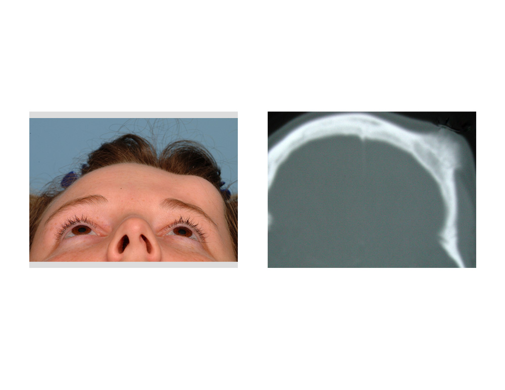 Case Study: This 34 year-old female had developed a low growing bump on her left upper forehead over the past three years. As it had been larger she began to develop sensitivity when pressing on it and more recent onset headaches. A CT scan showed an invasive osteoma that had penetrated beyond the outer cortex of the forehead bone but also down into the inner cortex with expansion on the intracranial surface.
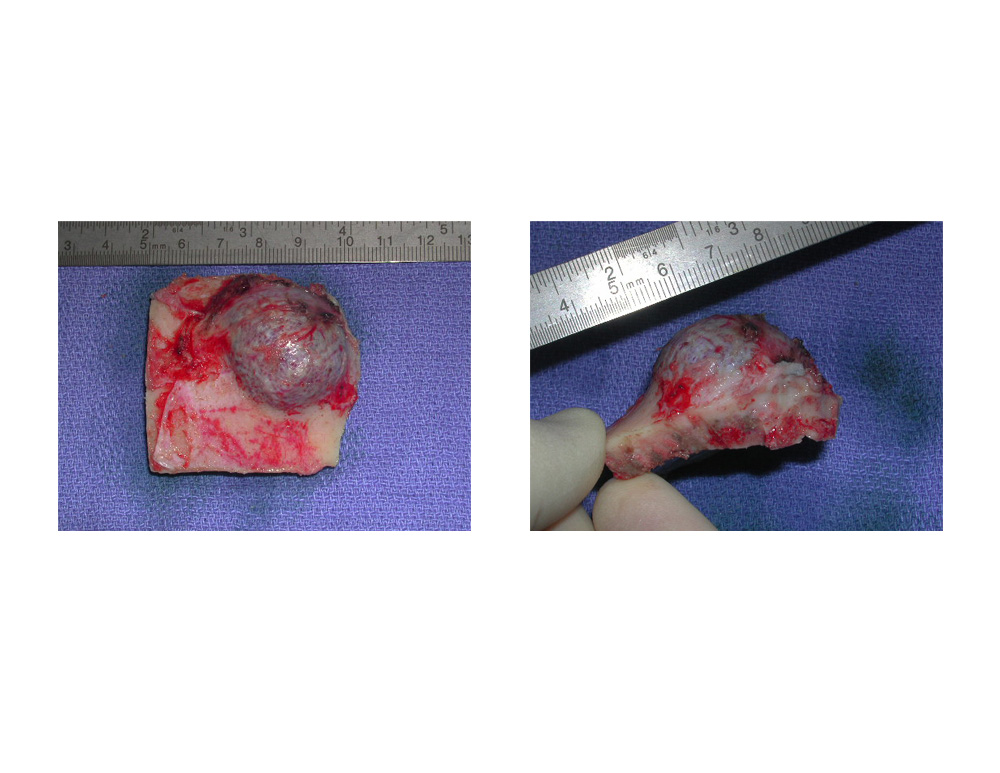
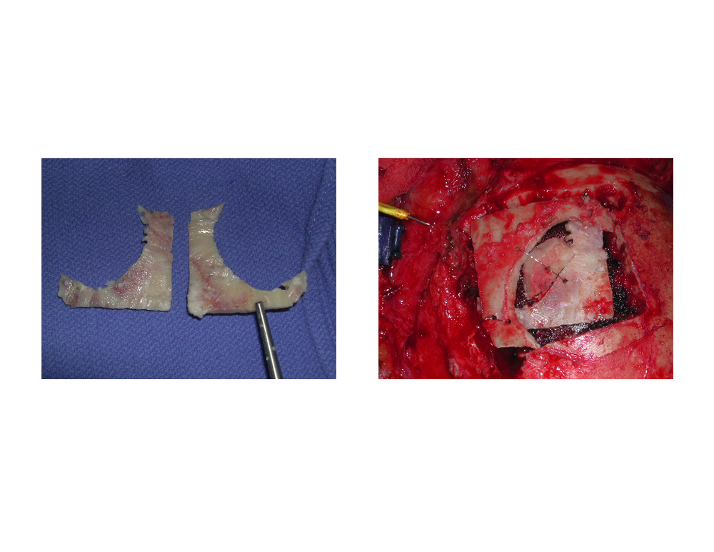 Under general anesthesia a bicoronal scalp incision was made to expose the entire forehead. A large frontal bone flap was cut and removed that incorporated the entire osteoma. The osteoma portion was removed and the remainder of the normal bone saved. This cranial bone segment was split into two pieces and was used to reconstruct a portion of the outer cortex of the forehead with plates and screws. To ensure optimal smoothness, the remaining bone defect not covered by bone with hydroxyapatite cement.
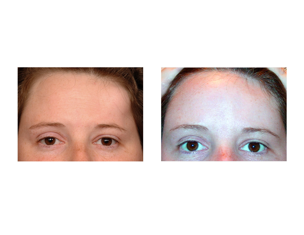
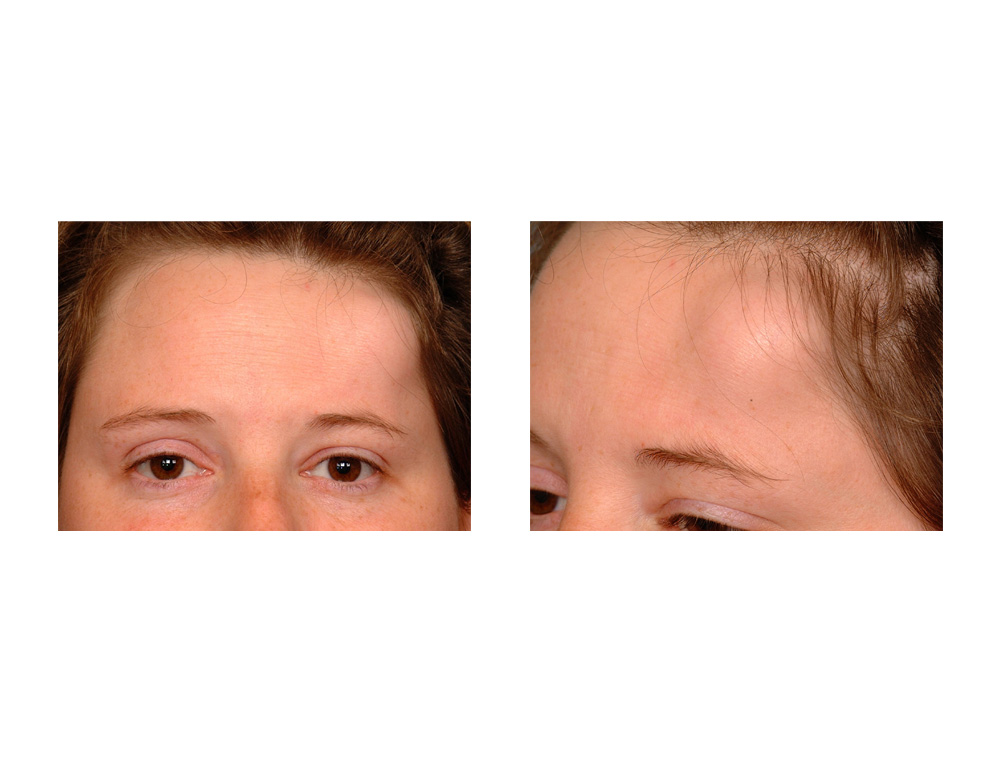
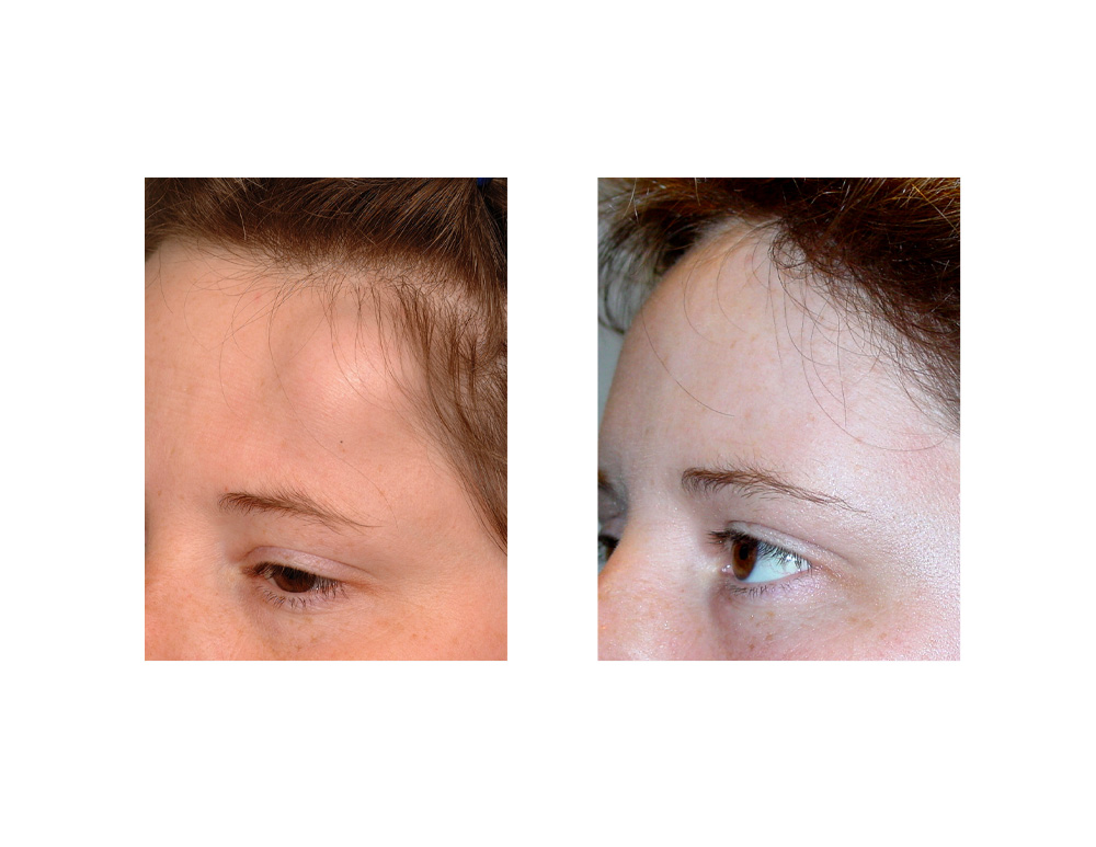 Three months after surgery she maintained a smooth and symmetric forehead contour. Most of the numbness of the forehead had resolved and she no longer had any headaches.
The vast majority of forehead osteomas as small, benign and easily removed by separating them from the surrounding bone. Large and more infiltrating osteomas are rare and can cause significant symptoms by their expansion of the persiosteum and even pressing on the dura. They require a full thickness bone flap to safely remove them from the underlying dura. Reconstruction can be done by a variety of skull restoration methods using bone grafts, alloplastic materials or a combination of both.
Case Highlights:
1) Large osteomas of the skull are uncommon but can be particularly deforming in the forehead and are often associated with pain. (headaches)
2) Full thickness resection of forehead osteomas requires a craniotomy flap for access.
3) Reconstruction of the resultant full-thickness forehead defect can be done by autologous bone grafts combined with contouring bone cement.
Dr. Barry Eppley
Indianapolis, Indiana