Background: The head has a wide variety of shapes and sizes. Like the face, there are certain head shapes that are more pleasing than others. While one knows intuitively whether they like their head shape or not, there are certain measurements of height and width of the head that can help classify its beauty or conversely its degree of deformity.
Head and face measurements and their ratios have been studied for over 100 years in a field of scientific study known as anthropometry. Classic anthropometric measurements of the head are its length, width and cephalic index. The length of the head (front to back) is measured from the midpoint of the brow just above the nose back to maximal projecting point of the back of the head. The width of the head is from a point just above the ears from one side to the other. Taken together the cephalic index is derived which is obtained by taking dividing the width of the head by its length which creates a percent ratio. This number is almost always less than 1 since most normal human skulls are longer than they are wide. Based on their cephalic index, head shapes have been historically divided into three main types; long-headed (dolichocephalic, > 80%), medium-headed (mesocephalic, 75% to 80%) and round-headed (brachycephalic, < 80%)
The dolichocpehalic head is one that has a narrow head width. (which is compensated for by an increased head length) But there are certain head shapes that are narrow in their bitemporal width but do not have an increased cranial length. Their mid-temporal region slants inward as it ascends upward to the top of the skull rather than having a more aesthetically pleasing convex shape on the side of the head.
To date, there has not been any known method to safely and easily create aesthetic augmentation for increasing the width of one’s head should their bitemporal width be too narrow.
Case Study: This 35 year-old young man did not like the narrow width of his head. He felt his head was too narrow above the ears and it slanted inward rather than outward. This made his head ‘too small’ and disproportionate for the rest of his head and face shape. He wanted a wider head but did not want any visible scars in doing so given his close cropped hair.


While he had some moderate temporal swelling after surgery, his pain was minimal. He had little recovery other than some swelling that resolved in a few weeks. His head width was instantly changed into a more convex shape which was very pleasing, adding 1.5 cms of bitemporal width. (Due to patient privacy, he did not want his before and after pictures published online. However he is willing to have them sent to anyone that wants to view them privately. You can request his before and afters by contacting me at inquiry@eppleyplasticsurgery.com)
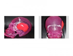
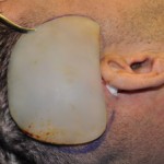
This type of temporal implants provide increased width and convexity for the narrow head. While custom temporal implants can be made from a patient’s 3D CT scan, the relative flat bony surface of the mid- and posterior temporal region makes a semi-custom approach a good treatment option. This new type of skull implant design provides another option in skull reshaping/augmentation that provides a different type of temporal augmentation that smaller more anterior-based implants for the non-hair bearing temporal hollow.
Case Highlights:
1) A narrow head is usually due to a bitemporal width reduction of the skull and/or muscle.
2) Custom temporal implants can be made to increase the bitemporal width from 5mm to 7mms per side.
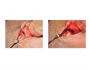
3) Large custom temporal implants can be discretely placed through incisions on the back of the ears.
Dr. Barry Eppley
Indianapolis, Indiana


