Background:The oral cavity is full of numerous bumps and protrusions, most of which are perfectly normal pieces of anatomy. The hard palate, however, is usually a completely smooth surface short of the small rugae or undulations of its mucosal covering. The actual hard palate is two pieces of perfectly smooth bone that comes together and unites in the middle very early in the formation of the fetus.
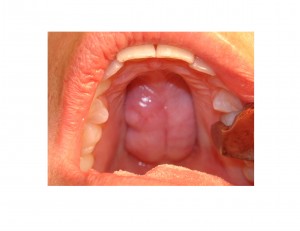
This smooth hard palatal surface can be dramatically disrupted by the presence of a hard bony bump or protrusion, commonly known as a palatal torus. These are bony growths that are usually present in the midline of the hard palate. While historically known as a torus, it is more accurately described as a benign osteoma. No one knows why they form or what causes them.For whatever reason they are much more common in women. While often less than 2cms in size, they can continue to growth throughout one’s life.
While palatal tori can be quite small and may appear as just a small and innocuous bump, they can become large. They can grow to the point of occupying much of the hard palatal surface. They can have numerous shapes from round to lobular. If they get big enough and are lobular is shape, they can cause numerous problems including food entrapment, irritation, ulcerations and difficulty with fabricating dentures.


Case Highlights:
1) Large palatal tori can cause symptoms, such as food entrapment and a source of irritation, and may necessitate removal.
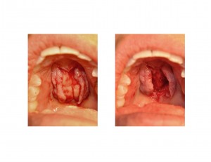
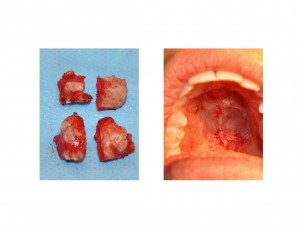
2) Palatal tori are surgically removed through a midline split approach separating the tori into multiple pieces with an osteotome.
3) Recovery after palatal tori surgery is minimal and they do not recur after their excision.
Dr. Barry Eppley
Indianapolis, Indiana


