

Background: While the ears are on the opposite sides of the head and can only be simultaneously viewed in the front view, this does not mean that people don’t care about ear asymmetries. The ears can be asymmetric for a lot of different reasons from how much they stick out to differences in their size and shape.
One of the ways ears can be different is in their length or vertical height. One ear can be longer than the other as measured from the top of the helix down to the bottom of the earlobe. Such height differences can be due to an increased amount of ear cartilage (a bigger cartilage framework) or a longer or bigger earlobe. That diagnostic difference determines whether a ‘high’ or ‘low’ vertlcal ear education should be done.
While the vertical height of the cartilaginous portion of the ear can be done by a wedge excision anywhere along its length to create a vertical reduction avoiding an unsightly scar for an aesthetic problem would be important. The most effective method to achieve a reduction and have a minimal adverse scar risk is the scaphal flap method. While this does cut across the helical rim, this is a very small section of the excision which places the scar on the inside of the helical rim where it is hidden.
Case Study: This male was bothered by a difference in the height of his ears. The right ear measured 65mm from the top of the curve of the superior helix down to the bottom of the earlobe. The left ear measured 70mms. The earlobes measured equal in their lengths so the difference in ear height was due to a longer cartilage framework
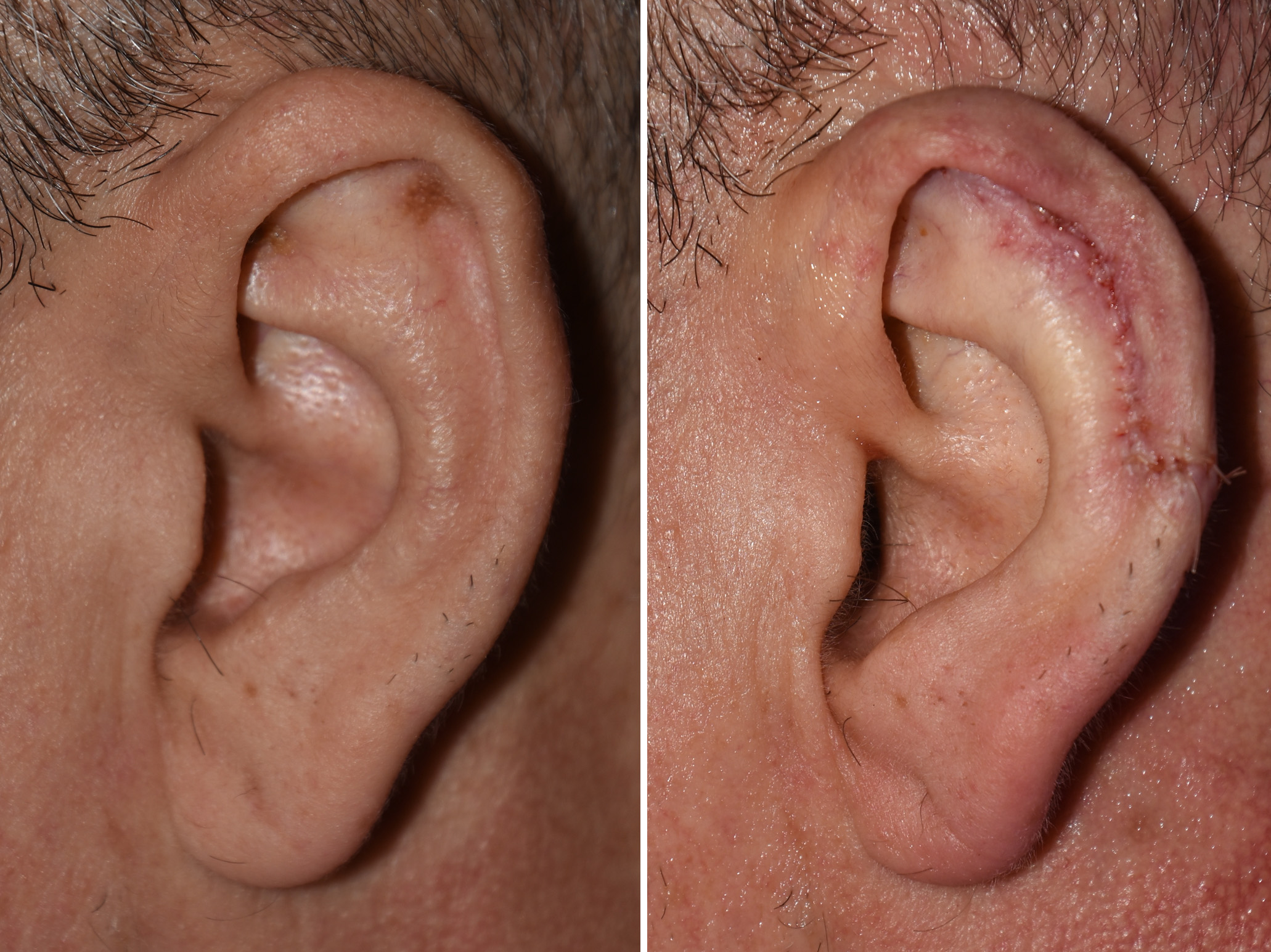 Under local anesthesia, a 5mm skin-cartilage excision was done through the outer helical rim and then up between the anti helix and helical rim where a more vertical wedge of front ear skin and cartilage was removed. The skin on the back of the ear was maintained. Closure was down by closing down the excised space by bringing down the superiorly pedicled cartilage-skin flap and putting together the cartilage and skin edges. In essence the 5mm vertical height reduction needed by transferred to a more limited back cut across the helical rim of the ear.
Vertical ear reduction with the scaphal flap method requires a bit of disassembly of the upper ear. This may seem ‘radical’ to those not experienced with its use but it is a very effective method that cleverly placed most of the scar inside the helical rim. But even the small scar that crosses over the helical rim heals well. Meticulous re-assembly of the cartilage and skin tissues avoids notching of the helical rim which is the only potential adverse scar issue with this ear flap technique.
Highlights:
1) One type of ear asymmetry is differences between their two sides in vertical length.
2) When the cause of the asymmetry is the cartilage framework, the most scarless method for vertical ear reduction is the scapula flap method.
3) Scaphal flap vertical ear reduction can be performed under local anesthesia.
Dr. Barry Eppley
Indianapolis, Indiana