Aging around the eyes occurs just like that of the rest of the face through loss of volume and thinning of the skin with wrinkle formation. While a variety of well known surgical techniques are available to rejuvenate the eye area (blepharoplasty and browlifts), the use of eye fat injections is still regarded as precarious by many surgeons. This is because fat injections are extremely prone to irregularities in the thin eyelid skin creating a high potential for an unsatisfactory aesthetic outcome.

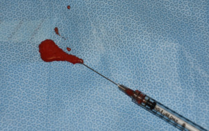
In the January/February 2016 issue of the JAMA Facial Plastic Surgery journal, an article was published entitled ‘Fluid Fat Injection for Volume Restoration and Skin Regeneration of the Periocular Aesthetic Unit’. In this paper the authors describe their techinque for injecting fat around the eyes. They have named their eye fat injection technique superficial enhanced fluid fat injection. (SEFFI) With this technique they do not process the fat but rely on the small size of the side ports of the harvesting cannula to use only small fat lobules immersed in a stromal component. This fat extract is then mixed with platelet-rich plasma. (PRP) This admixture contains viable fat cells, adipose-derived stem cells derived from the stromal vascular fraction and the platelets from the PRP. The concentrate of PRP makes up 10% to 20% of the total mixture to be injected. This creates a fine yellow paste-like mixture that flows through smaller needles in a more linear fashion which dramatically reduces the risk of lumps or irregularities.
In the video accompanying the article, it shows an injection of the SEFFI material flowing easily through a 23-gauge needle with the consistency of a synthetic hyaluronic acid filler. The SEFFI material is injected into the brow and upper eyelid sulcus above the eyelid crease including at the apex of the A-deformity at the medial third of the upper eyelid. The lower eyelid is approached from its lateral side to correct the temporal depression at this level, and it is extended along the infraorbital hollow, the eyelid-cheek junction, and the tear trough just below the medial canthal tendon. Most of these eyelid areas would be off limits with traditional fat grafting due to the high risk of fat irregularities.
It is clear that improving the use of eye fat injections requires particulation of the traditional fat graft. Liquifying the fat and adding PRP makes both biologic and technique sense when injecting into this sensitive facial area.
Dr. Barry Eppley
Indianapolis, Indiana


