Injectable fillers, along with Botox, have revolutionized the treatment of the aging face particularly one early in the process. The number of such injectable facial treatments around the world must surely number in the billions at this point in time over the past twenty years. The typical complications from injections are largely aesthetic and are well known. There are few major complications that have been reported, of which skin loss or necrosis is the most dire consequence.
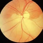
Irreversible vision loss can now be included in the list of major complications from injectable facial fillers. In the March 2014 issue for JAMA Ophthalmology, an article appeared entitled ‘Cosmetic Facial Fillers and Severe Vision Loss’. In this paper, three patients were reported that had central retinal artery occlusion right after receiving injections of different injectable facial fillers in the forehead area. All three patients had injections of either a hyaluronic acid filler, fat and Artefill in the high forehead area. Adverse changes to the retinal circulation were demonstrated by fluorescein angiography. All three patients had persistent visual field defects that did not resolve.
While there have been previous reports of eye complications from injections of other facial areas, this is the first report of it happening from injections into the forehead. While extremely rare, the highly vascularized tissues of the entire periorbital area make it an ever present albeit remote possibility.

According to the American Society of Plastic Surgeon’s statistics for 2013, well over two million injectable filler treatments were performed…and this number does not include every other type of doctor who may perform them. Even if these were the only cases of visual problems that have occurred in a single year, this places the risk at 1 in a million or more treatments. While rare, it can still happen and the best way to avoid it is to use microcannulas for injections and not needles. This makes it much harder to inadvertently enter the vessels around the eye.
Dr. Barry Eppley
Indianapolis, Indiana


