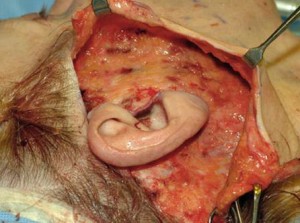
The classic teaching in facelift surgery to avoid injury to the greater auricular nerve is to identify the McKinney point. This point represents the location of the nerve trunk which is 6.5 cms below the ear canal on the sternocleidomastoid muscle. Further delineation of the nerve distribution was described by Ozturk with a 30 degree angle from the Frankfort horizontal plane which outlines the region of nerve distribution. Staying right under the skin and above the fascia over this area will avoid inadvertent nerve injury.
In the March 2017 issue of the journal Plastic and Reconstructive Surgery an article appeared entitled ‘What Is The Lobular Branch of the Great Auricular Nerve? Anatomical Description and Significance in Rhytidectomy’. In fifty cadaver dissection the lobular branch of the greater auricular nerve was dissected out. Various measuremenst were taken to the ear, SMAS and mastoid process. Greater auricular nerve diameter was measured. The branching pattern of the nerve and the location of the branches within the Ozturk 30 degree angle were documented.
The lobular branch existed in all specimens and was distributed to three regions. In the vast majority of the time (85%), the lobular branch was located inferior to the antitragus, in the remaining specimens it was located inferior to the tragus. The path of the lobular branch can be determined before surgery by making two vertical lines from the tragus and antitragus down to the McKinney point. The lobular branch ascends within this marked region. These markings provide guidance to avoid injuring the lobular branch during facelift flap dissection and SMAS elevation.
Dr. Barry Eppley
Indianapolis, Indiana


