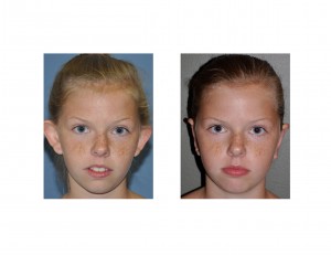
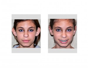
Even though the ears are located off to the side of the face, they can have an influence on one’s facial appearance. Being made up of a collection of ridges and grooves with a dangling earlobe hanging off of a cartilage framework, the ear is often not appreciated for its complex shape…until it is abnormal. The most common ear shape anomaly is when it sticks out too far.
The ear should not be a dominant structure on one’s head. With a geometry that is as individual as one’s fingerprint, it remains obscure as long as it sits at an angle of less than 30 degrees from the side of the head. Once going beyond this angle, the ear becomes too prominent.This prominence is caused by an alteration in the shape of the ear cartilage. The two most common causes of the protruding ear are absence of the antihelical fold and a large concha.


Many protruding ear problems are a combination of both a large concha and the lack of an antihelical fold. Often there may be a weak antihelical fold and a slightly large concha. The use of combined antihelical fold sutures and a conchal reduction/setback is often needed. These are the hardest protruding ear problems to treat and require a careful eye beforehand to make the diagnosis and intraoperative artistry and persistence to get the best ear shape.
One of the most overlooked problems in the protruding ear is the earlobe. Since it is not part of the cartilage structure of the ear, it will not lay back when the cartilage is reshaped if it is also protrusive. Often the earlobe must also be folded back by skin removal on the back of the earlobe as part of the otoplasty procedure.
In reshaping of the protruding ear, it is important to match the corrective technique with the cartilage shape problem. Failure to do so results in many of the postoperative otoplasty problems which consist of unnatural shapes and bends of the ear’s cartilages. Once the ear cartilages are resected, unnatural bends created, or set back too far, secondary correction can be difficult.
Dr. Barry Eppley
Indianapolis, Indiana


