


Background: The craniofacial skeleton is prone to have raised bony areas as well as depressions. (discrete convexities and concavities) The vast majority of these are normal skeletal contours or variations thereof. But occasionally a raised hard bump develops that occurs in an area that is normally smooth.
One such traditionally smooth craniofacial area is that of the forehead. While the forehead can have different shapes the one consistent feature that it has is a relatively smooth surface. As a result any hard bumps that appear become visible and violates the expected smooth surface appearance. One such common hard bump that can develop is an osteoma. This is a development of a round mound of bone whose pathology is believed to occur from a small perforating arterial vessel exiting from the bone. While trauma and bleeding in the subperiosteal space is a known osteoma cause, many forehead osteoma patients can not recall a specific traumatic event prior to its appearance.
The clinical diagnosis of a forehead osteoma is a fixed hard circular mass in which the skin slides back and forth over it. Most are less than 1 cm in diameter and 3 to 5mms in height. A CT scan can be done to verify the clinical diagnosis but that seems unnecessary in the vast majority of cases. Only in larger fixed bumps that have a more rapid growth history is a CT scan warranted to rule out abnormal bone growth that extends beyond the outer cortical bone layer.
Removing a forehead osteoma must balance the incisional tradeoff to do so. While the direct approach of an incision right over it is effective, most patients would prefer to avoid that visible scar location. (although if a deep horizontal wrinkle line exists over or right next to it, then the scar concerns may become irrelevant) Placing the incision at or just behind the frontal hairline provides adequate access in most cases for removal.
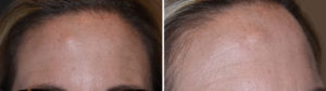 Case Study: This female developed a slow growing hard lump in her right upper forehead located 3.5cms below the hairline. It has been slowly rowing over the past few years and had become quite visible. It was painless and the skin easily move back and forth over it. She could recall no specific trauma to her forehead.
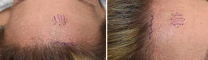
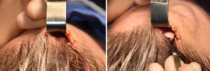
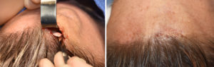 Under general anesthesia a 2.5cm irregular incision was made right at the edge of the frontal hairline directly above the osteoma. Subperiosteal dissection revealed a raised bony area that had a raised circular base. (like a mushroom) Osteotomes were used to remove it and smooth out the bone surface. Very small dissolvable sutures closed the neat invisible hairline incision.
Traumatic forehead osteomas do occur from a ruptured blood vessel and classically appear with a raised circular rim around the base…which is consistent with the vascular basis for their development in adults. The trauma that can cause this does not have to be so significant that it is memorable. These type of forehead osteomas are the easiest to remove with an osteotome because of their raised circular base. The use of an endoscope for their removal requires two scalp incisions to do so and offers a more ‘complicated’ technique that the direct hairline approach.
Case Highlights:
1) Forehead osteomas in adults often have a traumatic origin even if the patient does not have a specific recall of the event.
2) Raised circular-based forehead osteomas can be effectively removed with an osteotome technique.
3) The direct hairline approach to upper forehead osteomas provides direct visual access for their removal.
Dr. Barry Eppley
Indianapolis, Indiana