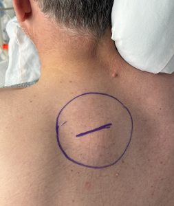
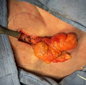
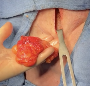
Background: Lipomas are a commonly occurring benign subcutaneous soft tissue tumor that occurs almost anywhere on the body. Many external appearing bumps or masses which feel soft can be presumed to be a lipoma. Once they appear they do tend to grow although such expansion is very slow and painless to the patient. A biopsy is rarely needed to make the diagnosis and they are usually diagnosed more by their appearance and history of growth. The same can be said for radiographic studies in which CT and MRI scans can make a clear diagnosis they often are not needed.
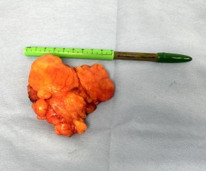
Lipomas have several unique features that distinguish them for other soft tissue tumors and are helpful in knowing to how properly remove them. First it is important to recognize that the external size of the lipoma is often just the ‘tip of the iceberg’. They always much bigger in size than they appear and, as a result, for certain sized lipomas removing them under general anesthesia is going to be more successful than trying it under local anesthesia. In many of the inadequately removed lipoma cases I have seen I am certain this was the reason. Secondly while this is an encapsulated mass that capsule is very thin and often not that distinguishable. What is distinguishable is the large fat lobules that are seen which are very different than that of the subcutaneous fat. You keep removing these large fat lobules until mall that is left or normal subcutaneous fat.
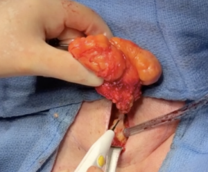
But the one very distinct feature that a lipomas has is its vascular pedicle. This is a benign tumor that has an artery and vein that feeds and drains it much like an organ. No matter how small a lipoma is its has a vascular pedicle. This must be found and cauterized to not only prevent any chance of regrowth bit also to prevent postoperative bleeding.
Case Study: This middle aged male has a long standing mass on the back of his left shoulder for almost twenty years. It slowly grew over the years until it got big enough that he started to resemble the ‘Hunchback of Norte Dame’.
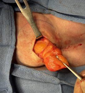
Closure was done with quilting sutures to avoid the need for a drain and restorable sutures for the skin in a subcuticular fashion.
As can be seen in this case the size of a lipoma on the inside is much bigger than its appears ion the outside. Thus complete removal may involve more surgery than one thinks.
Key Points:
1) Lipomas are always bigger tumors than they appear externally.
2) The pedicel of the lipoma must be identified and cauterized to prevent postoperative bleeding and recurrence.
3) The incision to remove a lipoma should be minimized in length and used a ‘mobile window’ in its removal.
Dr. Barry Eppley
World-Renowned Plastic Surgeon


