


Background: The most common ear reshaping procedure is a setback otoplasty for the correction of protruding ears. (technically earlobe repair is the most common ear surgery but this is not an ear reshaping surgery) In an otoplasty the ear generally has good components but is distorted by its protrusive condition.
In a much more uncommon aesthetic ear deformity is that of the large or long ear. (macrotia) This refers to the ear in which it is too vertically long from the very top of the superior helical rim down to the bottom of the earlobe. Normal ear heights are in the range of 50mms to 60mms depending on gender and physical size. In most cases of male macrotia that I have treated, vertical ear height is usually 65mms or greater. This can also be associated with an ear hat is also too wide in its upper third and with an earlobe that is too big.
The surgical treatment of macrotia is very different than that of the protruding ear. A setback otoplasty is a rearrangement of the cartilage shape of the ear while a macrotia reduction is about tissue removal. While a setback otoplasty achieves its effect through hidden incisions on the back of the ear, macrotia reduction must be done with incisions on the front of the ear.
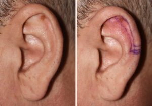 Case Study: This middle-aged male had long been bothered by his larger left ear. It was 6mms taller than that of the right ear with a larger scapha fossa of the upper third of the ear. The preoperative markings were made for a superiorly-based scaphal flap reduction technique.
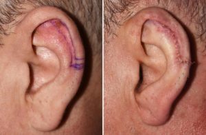
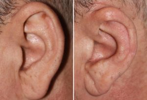 Under local anesthesia an anterior segment of skin and cartilage were removed with a 6mm backlit of tissue done across the mid-helical rim. This reduced the ear height by the desired 6mms. The immediate after surgery picture shows the result obtained and a comparison to a picture of the ear one year later shows how well the incisions has healed. Only a vert slight scar can be dine crossing the mid-helical rim on close inspection.
Despite the anterior location of these incisions, most of their length is placed on the inside of the helical rim where it is hidden. Only a very small length of the scar crosses the visible helical rim. The scar in this area is actually never a concern as it is s small. The development of any notching of the helical rim as it heals is what could potentially make it visible.
Case Highlights:
1) Vertical ear rejection requires the removal of ear tissue either from its northern or southern ends.
2) Excessive superior ear height often comes from an enlarged scapha fossa between the superior crus and the outer helical rim.
3) Superior-directed vertical ear reduction entails scapha fossa reduction.
Dr. Barry Eppley
Indianapolis, Indiana