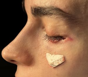
Background: Upper and lower jaw osteotomies, once known as orthognathic surgery, has undergone an evolution for what was its original purpose. Designed to correct dentofacial deformities for malocclusions, and while still used for that purpose today, its uses currently have expanded to the treatment of obstructive sleep apnea and as a method to increase mid- to lower facial projection for aesthetic purposes. One does not have to have a malocclusion to undergo the surgery and some patients do not even need orthodontic preparation for it.
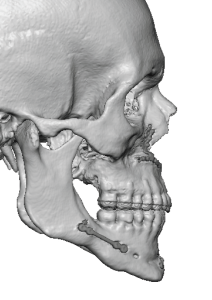
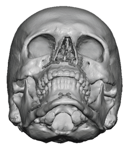
Now known as Bimaxllary (Bimax) Advancements the primary movements are forward and often significantly so. While these facial bone movements have many benefits they can create a facial imbalance or disproportion. The facial bones above the LeFort 1 level are left behind so to speak and this can result in cheek and lower eye (infraorbital) deficiencies which are seen as lack of cheek projection and undereye hollows.
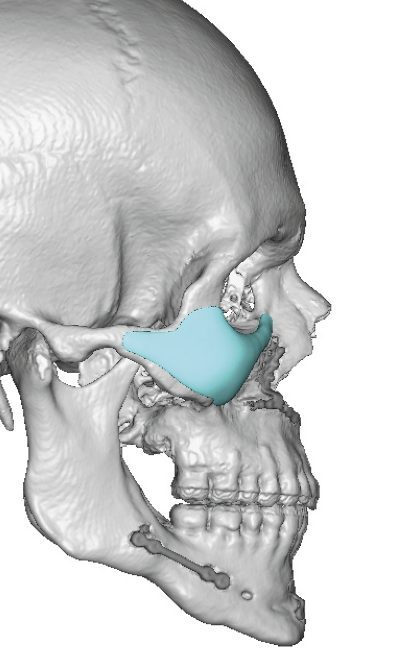
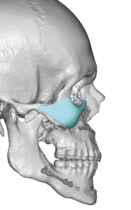
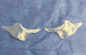
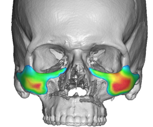
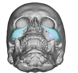
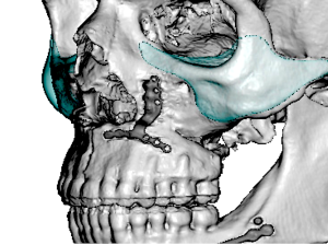
Unlike maxillary and mandibular bone movements through osteotomies there are no such anterior movement osteotomies for the orbits and cheeks. Augmentation can only be done by onlay augmentation of the bone with implants. And given the unique 3D shape of the orbits and cheeks custom designed implants are needed to achieve the proper effect.


Key Points:
1) Significant bimaxillary advancements leaves the cheeks and infraorbital rims behind which in some patients may create an unbalanced facial appearance.
2) Custom infraorbital-malar implants provide projection to the upper midface to bring it into better balance with what was achieved at the LeFort I bone level.
3) The lower eyelid is the best incisional approach to provide proper placement of upper nidface implant placement with the lowest risk of complications.
Dr. Barry Eppley
World-Renowned Plastic Surgeon

