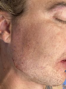
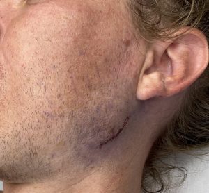
Background: Aesthetic manipulation of the jaw angle region, whether for implant placements or bone cutting work, involves elevation of the large enveloping masseter muscle. It is the insertion of the muscle along the inferior and posterior border that often must be elevated to perform the various procedures. The extent of this muscle elevation depends on the exact jaw angle surgery being performed.
The disruption of the insertion of the muscle would normally not be of aesthetic concern in these jaw angle surgeries unless it involves the placement of an implant. Since the implant acts like a spacer between the bone and the muscle, a physical block develops which can prevent the reattachment of the muscle in its original position. This becomes particularly relevant when an accidental disruption of the so-called ‘pterygomasseteric sling’ occurs.
The pterygomasseteric sling concept of the mandibular ramus is a misnomer. This implies that the masseter muscle on the outside and the pterygoid muscle on the inside of the ramus wrap around the edge of the bone and merge together. If so this would leave a thick layer of muscle that exists along inferior border of the jaw angle. In reality this does not exist. Each muscle insertion stops just short of the inferior edge of the bone. This leaves only a thin layer of periosteum along the inferior edge between the muscle on either side. The pterygomasseteric sling is a functional concept not an anatomic one.
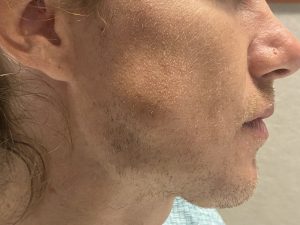
This is why in implant placement of the jaw angle region the risk of disrupting the musculoperiosteal masseteric attachment is not rare. Then when an implant is placed over the bone the muscle contracts superiorly having lost its inferior attachment. While this causes no functional issues it can create an aesthetic contour problem where the implant sticks out below the muscle. This becomes more apparent when the patient bites down, enlarging the detached muscle mass.
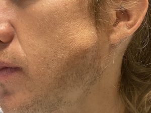
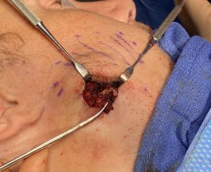
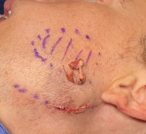
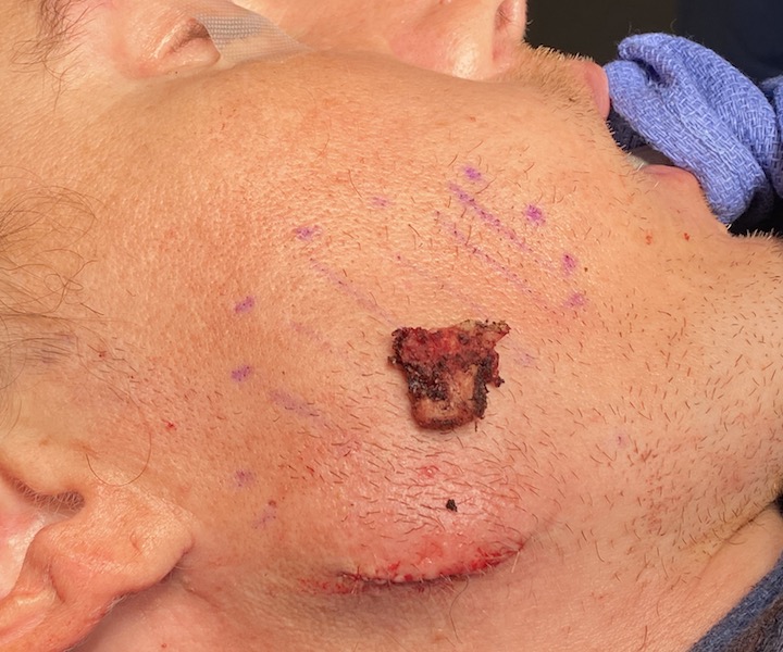
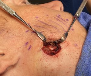
These unusually large masseteric muscle dehsicences were a combination of muscle contraction and heterotopic bone formation. The unusual amount of bone formation is a result of the periosteal reaction of the muscle. Most likely these were superior bone overgrowths from the prior implants that were never seen and removed from the implant surgeries. The transcutaneous approach provided good access for a satisfactory muscle repositioning over the jaw angle implants.
Case Highlights:
1) A direct transcutaneous approach can be used for masseteric muscle dehiscence repair.
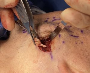
2) In this case of the treatment of masseteric muscle dehiscence after prior jaw angle implant surgeries, large masses of solid bone (osteomas) were discovered in the muscle.
3) The combination of intramuscular bone removal and masseteric muscle repositioning was able to be successfully done with the direct transcutaneous approach.
Dr. Barry Eppley
Indianapolis, Indiana

