

Background: The forehead is prone to the development of benign masses or growths more so than any other surface of the skull or scalp. Whether it is because it is more visible or whether it sustains more low level trauma the forehead is prone to developing benign slow growing masses. The most common forehead masses are osteomas and lipomas which represent the underlying layers of frontal bone and the subcutaneous fat of the scalp.
When diagnosing a forehead mass it can be very difficult to tell the difference by their appearance or feel. They both look round, creating a visible protuberance, and feel firm. Even a lipoma can feel firm. Neither one is mobile and each can feel like there is a rim around it. A CT scan will clearly make that differentiation if there is any reason to suspect any other diagnosis than osteoma or lipoma.
In removing benign forehead masses the two techniques are excision under direct vision and an endoscopic removal method. When referring to excision under direct vision I am not referring to making an incision right over the mass for removal. While that can be done most patients are not going to consider that approach very appealing. I am referring to making a more remote frontal hairline incision to do the excision. When to choose between a direct excisonal vs endoscopic removal method depends on where on the forehead it is located.
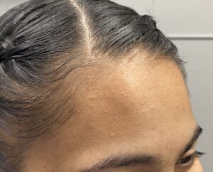 Case Study: This young female developed s right upper forehead mass that was slowly growing over the past several years. It was located in the upper half of the forehead, fell firm and immobile and an edge could be felt underneath it. It was presumed it was an osteoma. A preoperative CT scan was not obtained.
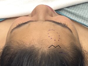
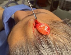 Under general anesthesia a small frontal hairline incision was made directly above the mass. In dissecting down to the mass it was quickly determined that it was not located in the subperiosteal plane but above it in the soft tissues. A lipomatous mass was seen and it was then dissected out as an encapsulated mass and brought out through the frontal hairline incision.
The technique to successfully remove benign forehead masses like osteomas and lipomas depends on where on the forehead they are located. If they are in the upper half of the visible forehead, as in this cases, it can be directly removed through a frontal hairline incision. If it is in the lower half of the forehead and closer to the brow bones an endoscopic is needed to guide the excision.
Case Highlights:
1) The most common pathologies for benign forehead masses are osteomas and lipomas.
2) Benign forehead masses if located in the upper half of the forehead can be removed under direct vision through small frontal hairline incisions.
3) An endoscopic removal technique for osteomas and lipomas becomes necessary when the mass is below the equator of the forehead.
Dr. Barry Eppley
World-Renowned Plastic Surgeon