

Background: Make chest enlargement or gynecomastia has a variety of different presentations. How much breast tissue enlargement, the amount of loose skin on the chest wall and the position of the nipple all influence the type of gynecomastia reduction technique that will be used.
Most gynecomastia techniques employ either direct excision by an areolar incision, liposuction or some combination of both. While effective for most young patients, the older gynecomastia patient often has one anatomic component that may change the surgical approach….nipple sagging or ptosis. This is defined very similarly to that of the female breast patient through the relationship of the nipple to the inframammary fold. And just like in the female when the nipple sits at or below the inframammary fold, no change in the breast/chest wall volume is going to make the nipple sit higher and look less ptotic.
In the older male gynecomastia patient, the nipples are often in a ptotic position. Even if the chest enlargement is not big, the nipple and its underlying breast tissue hand off the chest wall….creating an unattractive ‘man boob’. No amount of pectoral muscle development is going to lift the nipple up. And, in fact, significant pectoral muscle hypertrophy will make it look even worse by accentuating the overhang.
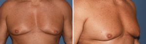 Case Study: This middle aged male, who had been an avid weight lifter/fitness enthusiast all of his adult life, was bothered by his nipple overhang. He had a large chest due to good pectoral muscle enlargement but the nipple ptosis was gradually getting worse. His options included trying liposuction and/or direct breast tissue excision initially to see how much improvement could occur. But I was not optimistic that this alone would provide any significant improvement in the nipple position on the chest wall. There was also concern that deflating the overhang may well make it look even worse. He opted for the one procedure that could guarantee an improvement in his nipple overhang.
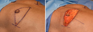 Preoperative marking included the location of the horizontal skin excision. Under general anesthesia the chest was initially treated by power-assisted liposuction to remove some overall volume. The horizontal skin excision was then done going just to the top of the areola, leaving an areola attached to the breast tissue behind.
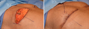
![]() The superior chest wall skin was elevated up almost to the clavicles. It was then advanced down over the lower chest tissues and areolar while it was being pushed and secured to the inframammary fold. The new nipple position was marked out using a triangulation method at 18 cms from the sternal notch, placing it just at the inferolateral pectoral muscle edge. The overlying skin was removed and the areolar derived up to the skin and closed.
The superior chest wall skin was elevated up almost to the clavicles. It was then advanced down over the lower chest tissues and areolar while it was being pushed and secured to the inframammary fold. The new nipple position was marked out using a triangulation method at 18 cms from the sternal notch, placing it just at the inferolateral pectoral muscle edge. The overlying skin was removed and the areolar derived up to the skin and closed.
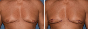
For significant nipple ptosis in the older gynecomastia patient, the nipple is going to have to be relocated. While free nipple grafting is always an option, there is the risk of depigmentation or portions of the nipple graft not surviving. Transposition of the nipple by keeping it attached to the chest tissues avoids these risks or at least significantly decreases them.
In solving the nipple overhang issue the trade-off is an inframammary scar. As long as it does not get into the sternum medially or extend to far out laterally, this is usually an acceptable aesthetic trade-off.
Case Highlights:
1) When nipple ptosis occurs in older gynecomastia males, no form of soft tissue reduction will elevate the nipples into a better or less ptotic position on the chest wall.
2) In such gynecomastia reductions the nipples need to be directly elevated by transposition or free nipple grafting.
3) In nipple transpositions setting its new elevated position. must avoid over correction or too high of a chest wall position.
Dr. Barry Eppley
Indianapolis, Indiana