

Background: The temporal artery is a well-known blood vessel located on the anterior side of the head. It is a branch of the external carotid artery that, when it reaches the temporal hair, splits or bifurcates into two separate smaller arteries. This anterior branch of the temporal artery, also called the superficial temporal artery (STA), is the only part of the temple artery that in some people can become visible. There are numerous reasons why the anterior branch can become visible from medical reasons such as arteritis and weight loss to that of trauma including injection injuries. But for the most part a prominent anterior branch of the temporal artery has no known reason for its occurrence.
For those who are bothered by the prominence of the temporal artery a ligation approach can be used to treat it. This approach is neither dangerous or risks any adverse effects are either the eye or overlying scalp hair growth. There is always been a considerable concern about these potential issues and, as a result, it is often told to patients that it should not be treated due to these potential dangers. But these are unfounded concerns and should not restrict those so interested from considering treatment.
While ligation is the only effective treatment for the prominent temporal artery this approach will fail if not properly performed. The most effective technique is to extract a loop of the artery at the various location points out through the skin incision. This not only helps keep the incision small but also ensures that a double ligation can be performed that is sufficiently apart. While single ligation done at each incision site may be sufficient there is no reason not to do it twice for absolute assurance of permanent cessation of flow through the vessel.
 Case Study: This male developed a right one-sided temporal artery prominence during COVID for no apparent reason. He had a vascular surgeon perform a single site ligation, who thought it was a vein, which did not work. No further treatment was recommended once it was discovered it was an artery because it was ‘too dangerous’ to do. In evaluating the prominence it had a classic arterial serpiginous course once leaving the temporal hairline. It had visible pulsatile flow to it.
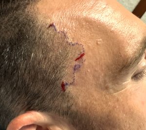 Its course was preoperatively marked out and three ligation incisions marked. (in red)
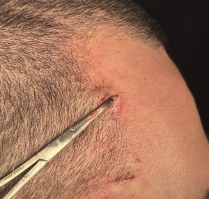
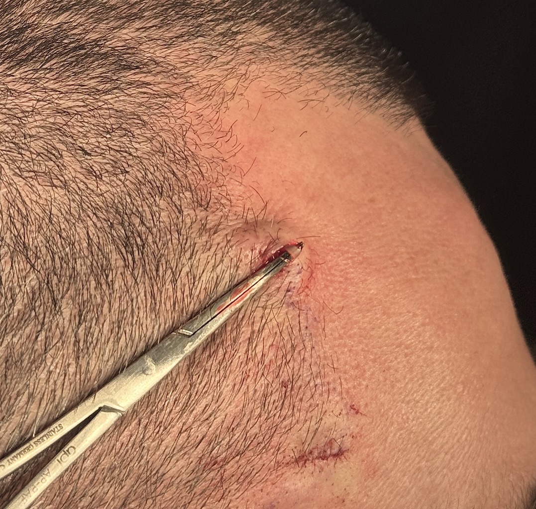
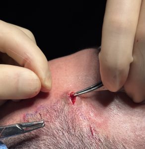 Under local anesthesia the three ligation sites were opened and the artery exposed. At each incisional site the arterial branch was pulled out of the incision making an external loop. Then double ligation was performed at the proximal and distal end of the loop with 5-0 Vicryl suture. The ligated loop was then dropped back inside the incision and the skin was then closed with running 6-0 plain suture.
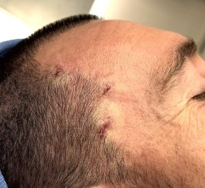 At the conclusion of the three ligations no visible, pulsatile or doppler pulse was evident along the course of the previously prominent artery.
Key Points:
1) The basic number of points for temporal artery ligations starts out at three and then pulsatile flow is evaluated.
2) Ligation locations are marked by the proximal location of the visible vessel from low anterior temporal hairline, the distal exit of the vessel into the scalp at the high hairline and the exposed vessel loop anterior to the mid-temporal hairline.
3) Temporal artery ligations are based on looped vessel extraction before ligation.
Dr. Barry Eppley
World-Renowned Plastic Surgeon