Background: Aging of the eyelids is always associated with varying amounts of skin excess, fat protrusion and skin wrinkling. The lower eyelid takes these issues to further ‘extremes’ creating additional aesthetic issues such as tear troughs, nasojugal grooves and a non-smooth surface contour in contrast to the underlying cheeks. These issues along with manipulating a suspensory structure stretched across the lower eyeball makes lower blepharoplasty surgery more challenging than upper blepharoplasty with higher risks of incomplete correction and eyelid positioning issues.
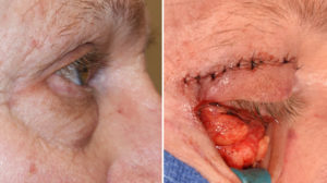
One anatomic feature that adds a level of complexity to lower blepharoplasty is the presence of a negative orbital vector. When the forward position of the cornea of the eye sticks out further than the infraorbital rim below it in side profile, this is known as a negative orbital vector. Its relevance to lower blepharoplasty is that it represents a bony deficiency of the infraorbital rim. This not only increases the risks of postoperative lower eyelid sagging (ectropion) but it makes creating a smooth lower eyelid contour more difficult as there is a bony deficiency as well contributing to the shape of the lower eyelid-cheek junction.
Augmentation of the infraorbital rim to add projection and support is the treatment method for the patient with the negative orbital vector who is to undergo a lower blepharoplasty procedure. What that augmentation method should be (fat, tissue grafts or implants) is open to surgeon and patient discussion of their various advantages and disadvantages.
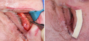
Case Study: This older male desired to improve his aging eye appearance. He had upper eyelid hooding and a large amount of herniated infraorbital fat protrusion and a deep indentation along the infraorbital rim. In side view he had a negative orbital vector which magnified the protrusive appearance of the fat. These maneuvers together with a conservative tapering 3mms skin excision across the lower eyelid incision created a smoother lower eyelid contour. The lower eyelids were closed by added support of lateral canthopexies and orbicularis muscle resuspensions.

Management of the lower blepharoplasty patient with a negative orbital vector must take into consideration the increased risk of postoperative ectropion and the contribution of lack of skeletal rim support to the eyelid’s shape. There are many different materials that can be used to augment the infraorbital rim and they all have their merits. For a more autogenous approach fat grafts and/or dermal implants are effective options.
Case Highlights:
1) The aging lower eyelid develops an exaggerated herniated fat appearance due to an infraorbital skeletal deficiency. (negative orbital vector)
2) Lower blepharoplasty in the face of a negative orbital vector manages both the fat excess and the deficient infraorbital rim.
3) One technique for infraorbital rim augmentation is allogeneic dermal grafts. (Alloderm)
Dr. Barry Eppley
Indianapolis, Indiana


