

Background
The temporal bone is the major skull bone located on the side of the head. Its primary functions are to protect the temporal lobe of the brain and encase the bony ear canal. Anatomically, it consists of two key regions:
-
A concave fossa located beside the eye, where most of the temporal muscle mass resides, and
-
A convex posterior portion that includes the ear canal and the bone above it.
While the majority of the temporal muscle volume lies within the concave fossa, its origin extends from the broad temporal line of the skull, sweeping downward into the fossa to attach to the lower jaw.
In temporal reduction surgery, performed for aesthetic purposes, the goal is to modify the side contour of the head from a convex to a straighter profile. This is achieved by removing the portion of the temporal muscle that overlies the convex part of the bone. Although this removed segment covers a larger surface area than the portion in the concave fossa, it contains significantly less volume due to its thinner structure. Consequently, there is no loss of lower jaw movement or function, even when the procedure is performed bilaterally.
The effectiveness of temporal reduction depends on the natural thickness of the temporal muscle, which is surprisingly substantial even over the convex bone area. The operation not only reduces the lateral fullness of the head but also alters its front-view profile from convex to straight. Since the bone itself has a slight outward curvature that the muscle accentuates, removal of this muscle flattens the contour.
Case Study
A male patient sought correction for a wide, convex-shaped head contour. On examination, he demonstrated a positive mouth-opening test—with wide mouth opening, the sides of his head appeared to narrow and straighten, confirming muscle-driven fullness.
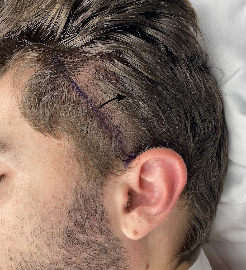
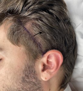 Under general anesthesia, bilateral postauricular incisions were made. The temporal muscle was carefully separated from both the overlying fascia and the underlying bone. A full-thickness, obliquely oriented muscle resection was performed, extending from the top of the ear upward to the bony temporal line. This incision defined the anterior resection line, while the muscle’s natural attachments delineated the superior, posterior, and inferior margins of the resection.
(In the corresponding picture, the purpose line is the muscle cut line internally and the back arrow indicates all muscle removed behind it)
The excised muscle segments were removed, and the fascia was meticulously reapproximated. A drain was placed, and the incision was closed within the ear crease. Fascia closure is a critical step, as it softens the transition created by the muscle resection and ensures a smoother contour.

 The head dressing and drains were removed the following day. Immediate improvement in the head’s shape and contour was evident. Equally importantly the patient exhibited no jaw range of motion linmitations.
Discussion
Although temporal muscle reduction is not widely known among patients—or even among many surgeons—it is a safe and effective method of reshaping the sides of the head. Despite concerns that such muscle removal might impair jaw function, no adverse effects on jaw movement have been observed in any cases to date.
Case Highlights
-
Temporal reduction reshapes the side of the head by removing excess temporal muscle.
-
The narrowing effect results from releasing and excising the muscle over the convex portion of the temporal bone.
-
Despite removing a large muscle surface area, no functional complications have been reported.
Dr. Barry Eppley
World-Renowned Plastic Surgeon