

Background: An aesthetic concern of the upper third of the face that has a vascular basis are prominent temporal arteries. Often confused as being veins prominent temporal arteries create an irregular serpiginous pathway across the temples into the forehead. Temporal veins do exist but they are straighter and look distinctly different. They can occur in conijntion with prominent arteries but more commonly they usually appear separately.
Prominent temporal arteries share numerous common features. They are not congenital in origin and develop later in life often after weight loss or facial fat loss due to aging. They are seen more frequently in men than women due to the larger walled arteries. They dilate and have increased visibility in response to heat, sunlight, exercise and alcohol. They can occur unilaterally but most of the time are seen own both sides of the temples. Often one side is more severely affected with a more visible material pattern than the opposite side.

 Having treated hundreds of prominent temporal arteries it has become apparent that there are some important anatomic differences in how they appear. Most commonly the takeoff of the primary feeding vessel is the anterior branch of the superficial temporal artery. This is a superior based or Type1 arterial prominence. In less common cases the primary feeding vessel or inflow from a branch off of the temporal artery in front of the ear. This is an inferiorly based or Type II arterial prominence. There usually is no evidence of a anterior branch of the superficial temporal artery. The differences between Type 1 and 2 arterial prominences are very visible and it will obviously change the proximal ligation location.

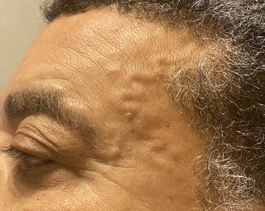
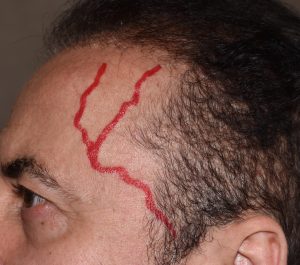
 Case Study: This older male developed very prominent temporal arteries after he lost weight. (20 lbs) They were more prominent on the left than the right but the arterial pattern was clearly a Type 2.
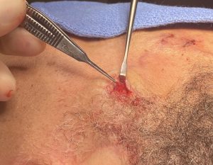
 Under local anesthesia proximal ligation points were done at the anterior edge of the sideburn supplemented by three superior ligation points at the elbow on the left and two superior ligation points on the right.

 At the completion of all ligation points a doppler is used to ensure that the arterial flow has been eliminated through the vessel pathway. If a signal is still heard anywhere along its course enough ligation points have not been done.
The difference between Type 1 and Type 2 temporal artery ligations is the location of the inflow. Most anatomic illustrations show the STA Y branch pattern of which the anterior branch is the feeder vessel as seem at the anterior edge of the temporal hairline. Less frequently the inflow comes from a branch off of the main trunk of the temporal artery located on front of the ear. This is rarely shown in anatomic diagrams but is easily visible. This changes the most proximal ligation point to a low rather than a high location
Key Points:
1) Type 2 temporal artery prominences have a primary feeder from a low accessory branch of the temporal artery.
2) Type 2 temporal artery ligations require a primary ligation T the anterior side burn area. (low temporal region)
3) Two to three high temporal ligations are needed at the ‘elbow’ of the arterial prominences.
4) Ligating until there is complete absence of a doppler signal along the course of the prominent artery is the key to optimal long term elimination of arterial show.
Dr. Barry Eppley
World-Renowned Plastic Surgeon