

Background: The scalp is pierced as a homogenous layer of soft tissue covering the skull whose most dominant feature is the hair it may or may not contain. While the scalp does have five distinct layers throughout its large surface area of coverage the thicknesses of it varies. The thickest part of the scalp is on the back of the head at the junction of the neck. Here the scalp crosses over the end of the visible occipital bone onto the attachments of the neck muscles.
Due to the action of the neck muscles through flexion and extension this creates a most mobile part of the scalp. As a result of this mobility and in certain male patients with thicker/shorter neck the scalp tissue bunches up. This creates a horizontal line of redundant scalp tissue known as an occipital roll. It usually lies along the nuchal ridge at the bottom of the occipital bone. An enlarged occipital knob (inion) or a raised nuchal bony ridge can be a cause of some occipital rolls in which the bone acts as a catchment point of the scalp tissue.
While single occipital rolls are the most common there are some patients that have double and even triple rolls on the back of their head. When two rolls exist the second roll may be above or below the primary roll over the nuchal ridge. But in the triple roll patient the 3rd roll is as a result of redundant neck tissue and is not really a true scalp roll.
The removal of an occipital scalp roll is excision with the understanding that the roll is being replaced by a fine line horizontal scalp scar. Despite having a scar as a tradeoff for roll removal it usually heals quite well and all patients to date have considered it preferable to the scalp roll. Two of the important keys to the occipital roll excision are incisional length control and avoiding injury to the greater occipital sensory nerve (stay above the deep fascia laterally on each side).
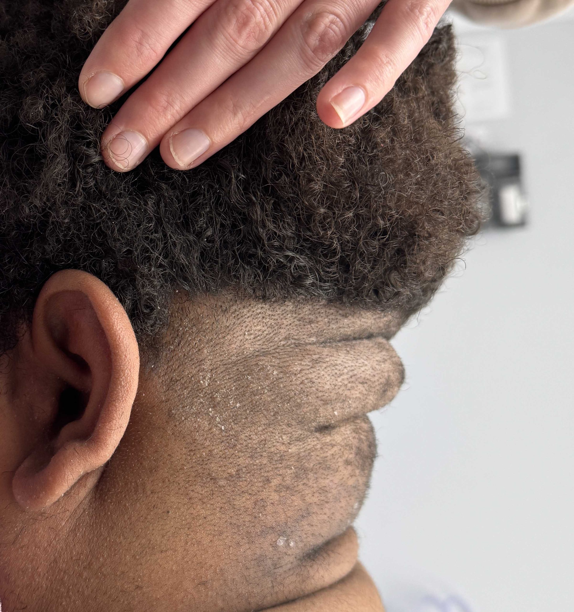
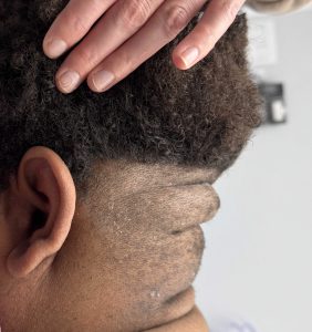 Case Study: This male had a large occipital scalp roll that was not visually evident due to his hair but was very palpable. When the hair was removed up above the roll it was very evident.
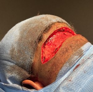
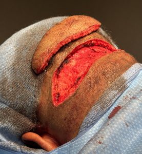 Under general anesthesia and in the prone position the scalp was excised horizontally above and below its perimeter/base. Centrally a wedge excision was done down to the bone which showed a minimally raised nuchal ridge which did not need to be shaved down. Laterally the excision was more superficial and did not extend down to the fascia to avoid nerve injury.
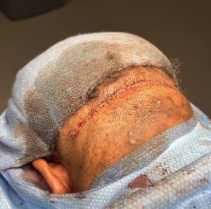 The lower neck tissues were the most flexible and, as a result, closure was done by advancing the neck flap superiorly for a multiple layer resorbable suture closure.

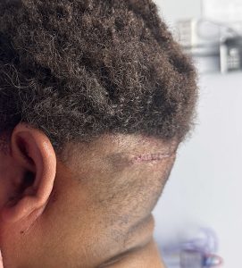 In side view the occipital roll was seen to be completely eliminated with a smoother skull to neck contour.
Occipital scalp rolls are almost always seen in males and have a tendency to occur in patients that have thicker or shorter necks. There is a variety of such roll manifestations in the number of roles that may appear. Most commonly hey single occipital scalp role is seen with a very small bunching of neck tissue beneath it. By the indented soft tissue indentations above and below it this provides an obvious elliptical incisional pattern with the location of the scar in a more natural location that is favorable for good scar healing. There has been a low incidence of the need for secondary scar revision although every patient should consider that as a possibility.
Key Points:
1) Thick scalps in males with broad necks have a tendency towards redundant soft tissue rolls tissues at the base of the skull at the back of head.
2) Such redundant scalp tissue can appear as a single, double or even triple horizontal rolls.
3) The occipital scalp roll if often associated with an enlarged central knob or nuchal ridges.
4) A horizontal excision is needed to flatten the prominent scalp roll with a fine line scar tradeoff.
Dr. Barry Eppley
World-Renowned Plastic Surgeon