

Background: Osteomas are well known benign bony growths that have a predilection for the skull. They are probably best known in the forehead as this prominent location drives patients to have them removed due to their visibility. They always occur as solitary bony bumps of varying sizes and quite frequently have a prior history of trauma. They are painless and can occur anywhere on the forehead but usually not on or near the brow bone.
Forehead osteomas in my experience can be either pedunculated or non-pedunculate which refers to their base. In a pedunculated osteoma its base is narrow and the bony growth emerges from it like a mushroom. In essence the base is more narrow than the osteoma itself. These are usually small and discrete in size. In non-pedunculated osteomas its base is its widest part and looks more like an expansion of the entire outer cortical plate of the frontal bone. They are usually larger in diameter and have more of an external mass effect.
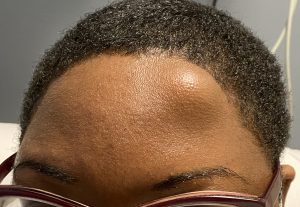 Case Study: This female presented with a large left forehead osteoma that had been present for years. She had a history of trauma to the forehead prior to its appearance. A CT scan showed it was a solid mass of bone that was restricted to an outcropping of the outer cortical table of the frontal bone. There was non inner table bone expansion.
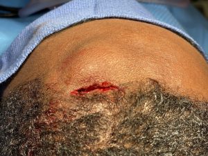
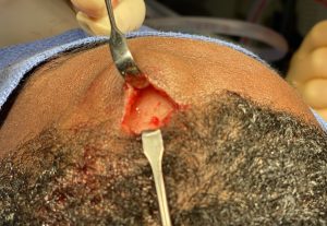

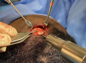
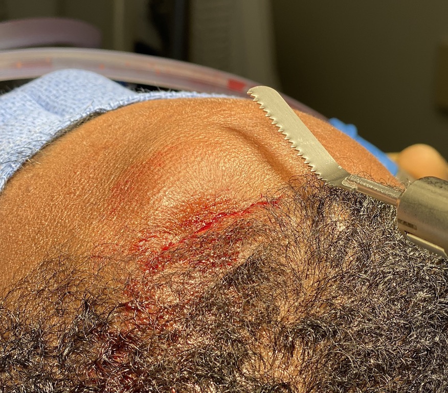
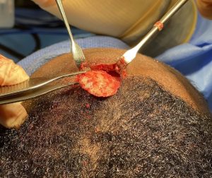 Under general anesthesia a 3 cm hairline incision was made directly above the osteoma and the bone mass exposed. An angled saw blade was used to cut across the base of the osteoma at the level of the surrounding normal forehead bone. Most of the bony mass was removed in one piece.
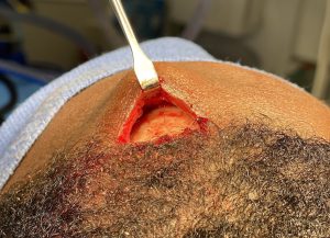
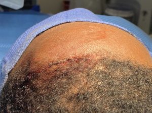 A high speed handpiece and burr was used to smooth out the base of the excision and the incision closed with resorbable sutures. A circumferential heads wrap was applied for overnite.
While most pedunculated osteomas can be removed with an osteoma due to their narrow base, pedunculate osteomas require power instrumentation to remove. A saw blade excision technique works well for them and some angulation of the blade is frequently necessary to make the cut level with the surrounding bone.
Case Highlights:
1) Larger forehead osteomas frequently result for prior trauma and have a wide non-pedunculated base.
2) One method for wide base forehead osteoma removal is saw blade excision through a hairline incision.
3) Saw blade excision is a safe and efficient technique that can be done in 30 minutes as an outpatient procedure.
Dr. Barry Eppley
Indianapolis, Indiana