

Background: Osteomas are well known benign bony overgrowths that frequently occur on the skull. The most common patient that presents for osteoma removal is when it occurs on the forehead for the obvious visibility reasons. It is easy to forget that the forehead (frontal bone) is part of the skull which explains their common occurrence there.
While is not so easily explainable is why forehead osteomas occur. Since trauma is the one reason that is known to be associated with the development of an osteoma it is hypothesized they have a vascular origin. Since the skull bone is replete with perforating vessels from the bone’s surface the purported mechanism is that a blood vessel ruptures which causes a small collection of blood under the periosteum. Since the periosteum is potentially osteogenic the blood serves as a nidus for bone formation. This then turns into a raised mound of slow growing bone that is circular in shape. It presents as a fixed hard mass in which the skin slides easily over it.
Regardless of its origin osteomas are removed by a sharp separation of it from the surface of the bone in which it usually separates uneventfully. The only question, and an aesthetically important one, is incisional access. A direct incision right over to can always be done but this is often unacceptable particularly in women. An endoscopic technique keeps the visible forehead free of any disfiguring scars.
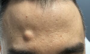
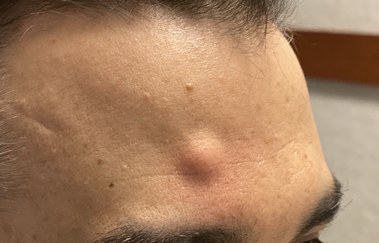
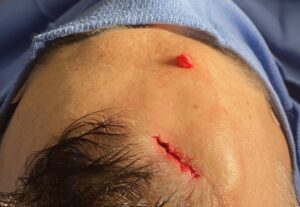
 Case Study: This adult male presented with a prominent hard fixed mass several centimeters above the right eyebrow. It was located on the lower third of the forehead.
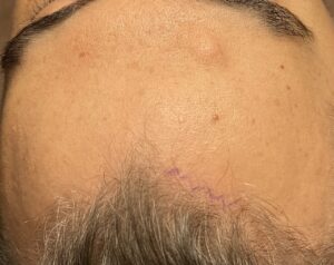
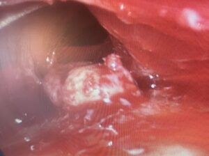 Under general anesthesia and using a single port endoscopic technique through a 2.5cm frontal hairline incision, the osteoma was visualized.
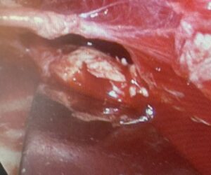
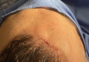
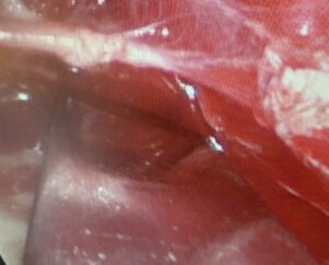 It was then removed with an osteotome. It separated cleanly leaving a smooth forehead surface behind.
 Forehead osteomas requires an endoscopic technique when they reside in the middle or lower half of the forehead. if they lie above the median horizontal line of the forehead they can be removed with an osteotome using direct visualization. When an endoscopic technique is needed I find that a single port works just fine even though the conventional forehead endoscopic technique uses two ports.
Case Highlights:
1) The most common technique for forehead osteoma removal is an endoscopic assisted method.
2) A single port endoscopic technique with an osteotome will work for most forehead osteomas.
3) Removal of forehead osteomas is not associated with a significant rate of recurrence