

Background: The temporal artery is best known in plastic surgery for its contribution in reconstruction as the vascular supply for local tissue flaps. The goal in these procedures is preservation of the blood flow to the artery to keep the transferred tissues alive. Conversely the strong blood flow in the temporal artery can have an aesthetic negative effect as it can make its serpiginous pathway into the forehead visible.
In treating many prominent temporal arteries I have observed that they consist of two types regardless of gender. These types differ in their visible pathway into the forehead based on which branch is enlarged. These different arterial pathways affect how many ligation points are needed.
The most common type of prominent temporal artery, which I will call Type 1, comes from the enlarged anterior branch of the superficial temporal artery. (STA) This pathway is well known and appears in any anatomic drawing or cadaveric dissection. It is easily diagnosed as it leaves the temporal hairline on the side of the forehead, becomes tortuous at it approaches the bony temporal line and then turns at 90 degrees to head up into the frontal scalp. When hair is not present even the Y configuration from the anterior branch emanates can become visible.

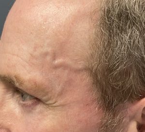 Case Study: This middle-aged male developed prominent temporal arteries for unknown reasons. He had no history of trauma or headaches associated with them. The right side was more prominent than the left but only slightly so. They had a strong palpable pulse in them that could be visibly along its entire visible course into the forehead.
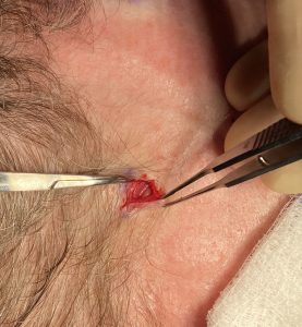
 Under local anesthesia three ligations points were marked and the ligations done in a proximal to distal direction. Through small incisions the artery was dissected out and pulled out of the incision to be ligated beginning with the one at the temporal hairline.
 Not unsurprisingly the most proximal ligation did not eliminate the arterial signal. The middle ligation area is where it makes a 90m degree turn north and a small incision is made in a wrinkle line closest to it when the patient raises their eyebrow.
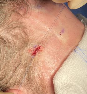
The final ligation point is done at the last visible extent of the artery before it heads into the scalp hairline.

 With three ligation points the arterial signal along the pathway of the artery was eliminated.
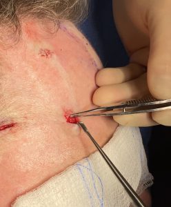
The six location sites were then closed with small resorbable sutures and the four behind the hairline were covered with small flesh-colored tapes.
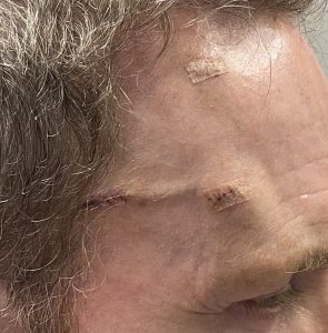
 When seen the next day the most prominent part of artery could be seen but it was non-pulsatile. Over the next few weeks the appearance of the artery will fade as the loss of blood flow will cause its diameter to fully collapse.
Case Highlights:
1) Prominent temporal arteries can arise from either the superior anterior branch of the superficial temporal artery (STA) or from the inferior frontal branch off the main trunk of the temporal artery.
2) Surgical treatment of the prominent anterior trunk of the STA may require as few as 3 ligation areas.
3) The definitive test of elimination of the prominent temporal artery is the elimination of the arterial doppler signal.
Dr. Barry Eppley
World-Renowned Plastic Surgeon