

Background: While there are a variety of pathologic conditions that can affect the scalp none is more unusual that cutis verticis gyrata. (C VG) While most people have seen someone so affected few realize that it is an actual pathologic problem. Appearing as wrinkles on the head is why is called gyrata being similar to the appearance of the surface of the brain. In most patients these wrinkle lines are not random but rather vertical running from front to back. (hence the term verticis or vertical) There can be single or multiple lines in the scalp and they can be slight in depth or very deep.
CVG is not present at birth and develops later in life. What causes it is not known although it appears somewhat similar to a facial condition known as linear scleroderma (LS) in terms of appearance, timing of onset and progression The difference is that LS is an atrophic condition of the skin and subcutaneous fat along vertical lines the following the path of the trigeminal nerve. CVG is a fibrosis/contracture of the skin and subcutaneous fat that, while usually vertical, is random in its scalp location although most occur on the top and sides of the head. Usually they occur one both sides of the head and are often perfectly symmetrical. (e.g., if there are three lines on one side there are three lines on the opposite side)
The historic method of treating CVG is excision of the indented scalp lines. While such excision is the only effective treatment for very deep V-shaped CVG lines, the scar tradeoff is less deep lines may not be acceptable or impractical when multiple lines are close together. An alternative treatment is release and fat grafting in the properly selected patient..
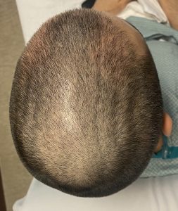
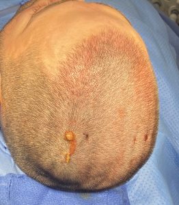
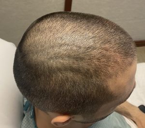 Case Study: This male had CVG appear after college as three shallow vertical lines in the scalp on each side. He was well aware of the condition as both of his brothers had it and it was known to be progressive. While he had hair coverage he preferred to wear it short where the lines were more visible.

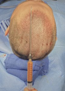
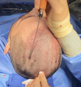 Under general anesthesia the linear scalp lines were initially injected with local anesthesia with epinephrine. Fat was harvested from his abdomen and flanks. Using a small stab incision in the middle of the line, a special ‘picklefork’ instrument that is a hollow cannula is introduced through the stab incisions and run under the scalp skin to release the indented lines. Because the skull is curved and the scalp lines follow it and the instrument is straight, approaching it from the middle in both directions allowed the subnormal release to be more uniform in depth.
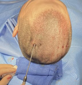
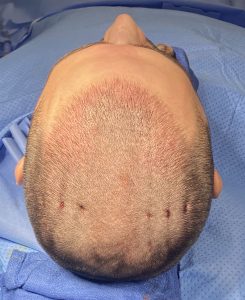
 Once the CVG is fully released fat is injected through the cannula by a syringe attached to it that contains concentrated fat from the abdominal/flank harvest. Enough fat is injected so that when the cannula is removed some fat will come back out. A single suture in each stab incision helps contain the injected fat.
 In very deep CVG in which the skin is very fibrotic an adequate release can not be done and excision would be the only treatment option. But in less deep CVG or early onset CVG excision is too aggressive for the problem. While it is not yet known how effective release and fat grafting is for CVG (not enough patients have yet been done), its autologous nature has almost no risk and is anatomically sound given the loss of the deeper tissues of the scalp under the lines.
Case Highlights:
1) CVG appears as linear fibrotic lines in the hair bearing part of the scalp whose etiology is unknown.
2) One treatment option its subcuticular release with injection fat grafting using a specialized instrument to do so.
3) It is not yet known how effective this treatment is for CVG or whether it will stop its progression but it is a benign treatment using the patient’s own tissues so there is little downside to it.
Dr. Barry Eppley
Indianapolis, Indiana