

Background: Facial asymmetry has highly variable presentations. But in many cases a smaller jawline on the affected side is seen. While this is typically perceived as a smaller jaw angle area, closer inspection by 3D CT scan analysis will usually reveal that the entire jawline is deficient from the chin posteriorly back to the jaw angles.
In days past the common approach for jawline asymmetry correction (that did not involve a malocclusion) was to ‘eyeball’ the problem. The surgeon would take some standard implants, blocks or sheets and hand made what looked like a symmetric correction to the opposite side. While often looking correct on the operative table, longer term postoperative followup often revealed an either inadequate correction, an over correction or the appearance of a new form of jawline asymmetry. Rarely was an optimal correction obtained by a visual intraoperative method.
Today the use of 3D CT scanning technology has left ‘eyeballing’ facial asymmetry problems a technique of the past. Measurements on the 3D CT scan or the analysis of printed out anatomic models allows the planning of jawline asymmetry correction to be done before surgery. This leaves the main task of surgery to place preoperatively designed implants as they were designed on the models.
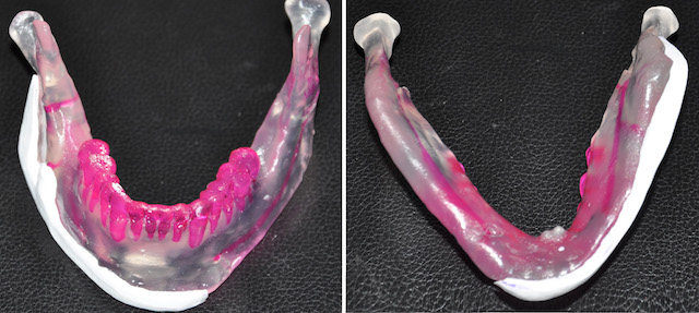
 Case Study: This young male had a history of a right subcondylar jaw fracture as a child. It was treated by some form of intraoral open reduction technique. As he continued to grow through his teenage years he developed a right facial asymmetry with a smaller jawline on the original fractured side. A 3D CT scan had done from which a printed anatomic model was made. This showed the healed but collapsed condylar segment on the right side with a shorter and smaller ramus.

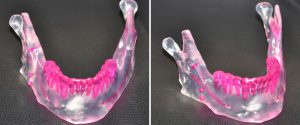
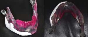 Using the anatomic model, a jawline implant was designed to match they shape of the opposite jawline. It could be seen to extend the whole way from the chin back to the jaw angle. An extension of the implant design was made up towards the oblique line back by the molars for the intraoperative placement of screw fixation.
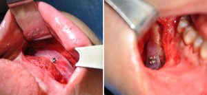 Under general anesthesia and through an intraoral anterior and posterior vestibular incisions, the jawline implant was placed in a subperiosteal pocket along the inferior border. Small screw fixation was done both at the anterior and posterior sections of the implant.
While this jawline asymmetry case used an anatomic model to fabricate the implant, today this would be done ‘model less’ using 3D CT designing software technology. This would provide an exact mirror image of the opposite jawline with much less fabrication time.
Case Highlights:
1) Jawline asymmetry often involves the entire side of the jawline not just the jaw angle area only.
2) Anatomic models are one method of creating a custom implant model for jawline asymmetry.
3) Most jawline asymmetry implants are quite thin but extend from the chin back to the jaw angles.
Dr. Barry Eppley
Indianapolis, Indiana