

Background
Injectable fillers are among the most commonly performed nonsurgical aesthetic procedures for the face, having been used hundreds of millions of times over the past two decades. While a wide variety of filling agents exist, all share one essential aspect: they must be injected.
Traditionally, this was done using a beveled needle to puncture the skin and reach the deeper subcutaneous tissues. More recently, blunt-tipped cannulas have become more commonly used. However, even these require a sharp needle to first traverse the dermis before the cannula can be introduced.
Among the potential complications of injectable fillers, intravascular injection leading to embolization and subsequent tissue necrosis is the most feared. This risk is heightened in the central face, particularly around the nasolabial folds, due to the proximity of the facial artery as it ascends along the side of the nose. In addition to embolization, another potential but less recognized complication is arterial wall injury, which can result in the formation of an aneurysm.
Facial artery aneurysms appear as pulsating bulges visible on the skin’s surface. While typically not painful, they pose a significant aesthetic concern and may enlarge over time, becoming more prominent.
Treatment Considerations
The only effective treatment for a facial artery aneurysm is ligation or ligation with resection. This involves a direct incisional approach to isolate the aneurysm, ligate its arterial branches, and excise the dilated segment to eliminate the mass effect.
Case Study
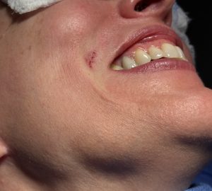
A female patient presented with a persistent pulsating bulge at the lower end of her nasolabial fold, just lateral to the mouth corner. This abnormality had developed following injectable filler treatment performed 17 years earlier. Multiple providers had declined to treat the lesion, citing concerns over the risk of nasal skin necrosis if the facial artery was ligated.
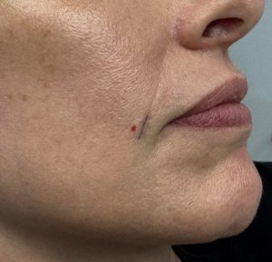
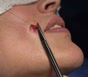
 On examination, the pulsatile mass corresponded to the red dot in the diagram. A strong Doppler signal confirmed its arterial origin. An incision was made along the nasolabial skin crease (indicated by a purple line in the original diagram), just medial to the lesion. Through a 6mm skin incision, an arterial loop was identified and dissected. Part of the artery was visibly enlarged.
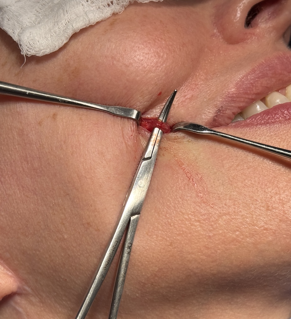
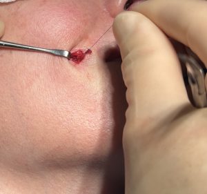 Further dissection revealed the bifurcation of the facial artery, with its characteristic Y-shape. Ligation sutures were placed at each of the three branches, and the aneurysmal segment was excised.
 The skin was closed using fine resorbable sutures. The previously strong Doppler signal was absent, confirming successful vascular isolation.
Discussion
Facial artery aneurysms are rare. In prior cases I have treated, no definitive etiology could be identified. In contrast, this case presents a clear and specific cause: needle injury during injectable filler treatment, leading to localized arterial wall damage and aneurysm formation.
There is a commonly cited fear that ligating the facial artery can cause ischemia or necrosis of nasal tissue. However, this concern stems from documented cases of intra-arterial filler embolization, where distal capillary occlusion and limited collateral flow led to nasal skin loss.
In this case, ligation of the ascending branch of the facial artery on one side did not compromise nasal skin perfusion. Adequate cross-flow from contralateral anastomotic connections maintained healthy vascular supply.
Key Points
- Facial artery aneurysms are rare but can be caused by needle trauma during injectable filler treatment.
- This case illustrates an aneurysm that developed at the bifurcation of the facial artery, presenting as a pulsatile bulge 17 years after injection.
- Treatment through a small, well-concealed incision allowed for precise dissection, ligation, and excision of the aneurysm with excellent vascular and aesthetic outcomes.
Dr. Barry Eppley
World-Renowned Plastic Surgeon