

Otoplasty correction of the protruding ear can be one of most satisfying plastic surgery procedures one can undergo. The immediate change of prominent ears that stick out to an instantaneous normal appearance is often nothing short of dramatic. While the cartilaginous cause of the prominent ear can be easily determined, its correction involves a lot of artistic technique in its reshaping. As a result, undesireable otoplasty results can occasionally happen as the surgery is not an exact science.
In the August 2011 issue of Plastic and Reconstructive surgery, a good article on the secondary correction of the unfavorable result after otoplasty was published. The article covers both the secondary treatment of undercorrection and overcorrection. The section on undoing overcorrection of an otoplasty (ears pinned back too far) interested me the most as this is by far the more difficult problem.
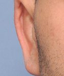
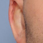
While often lumped into the name, ‘telephone ear deformity’, ear overcorrections are apparent as the outer aspect of the ear touches or nearly touches the side of the head. This makes the outer edge of the concha being the visible outer edge of the ear rather than the helical rim. This could happen because the sutures used to create the antihelical fold are pulled too tight or back too far, the conchal hypertrophy cause of the prominence is left untreated, or too much skin has been removed from the back of the ear.
Ear overcorrections essentially take an ear, which has too much tissue or cartilage that is abnormally shaped, and turns it into a situation of relative tissue deficiency. As the authors point out in their revision description, the ear needs to be released from a postauricular approach and can often reveal a skin deficiency and a need for cartilage grafts. These two issues must be taken into consideration before surgery. If skin on the back of the ear is deficient, either a sliding skin flap from a sulcus incision or ‘finger flaps’ from the mastoid must be used to replace the missing skin. The donor site will require a small skin graft.
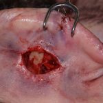 Once the ear cartilage is released, cartilage grafts will be needed to stent open a collapsed antihelical fold or to reinforce areas of cartilage fracture that have occurred from the release. Such cartilage grafts can only come from two sources, the ear concha itself or a small rib graft. The choice will be based on how much cartilage reinforcement is needed.
Reconstructing an overcorrected otoplasty , also known as a reverse otoplasty, can be difficult but almost always needs tissue grafts. If an overcorrection is treated very early after the initial otoplasty, suture release alone may be sufficient. But once scar and tissue adhesions is established, months to years later, the issue becomes of one of cartilage release and reinforcement and skin flaps or grafts.
Dr. Barry Eppley
Indianapolis, Indiana