

Avoiding injury to the frontal (temporal) branch of the facial nerve is the key safety issue during cosmetic superficial temporal artery (STA) ligation. The protection strategy is primarily anatomic plane control and incision placement.
1. Know the Nerve’s Course
The frontal branch travels:
- From the parotid gland
- Across the zygomatic arch
- Toward the lateral brow and frontalis muscle
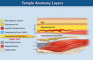 It runs within the superficial musculoaponeurotic system (SMAS)/temporoparietal fascia layer, just deep to the subcutaneous tissue in the temple.
A commonly used landmark is Pitanguy’s line:
0.5 cm below the tragus ? 1.5 cm above the lateral eyebrow
The nerve generally runs along or just below this line.
2. Safe Dissection Plane
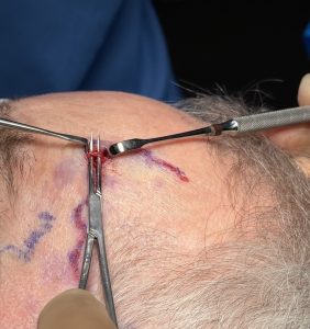
 The potential risk of nerve injury is in the distal ligation sites out in the exposed skin not the one at the hairline. For cosmetic artery ligation, stay superficial to the temporoparietal fascia.
Safe plane
- Skin
- Subcutaneous fat
- Superficial temporal artery
Danger plane
- Temporoparietal fascia / SMAS
- Where the frontal nerve branch runs
Key principle:
 Dissect in the subcutaneous layer only.
Do not enter the SMAS/temporoparietal fascia.
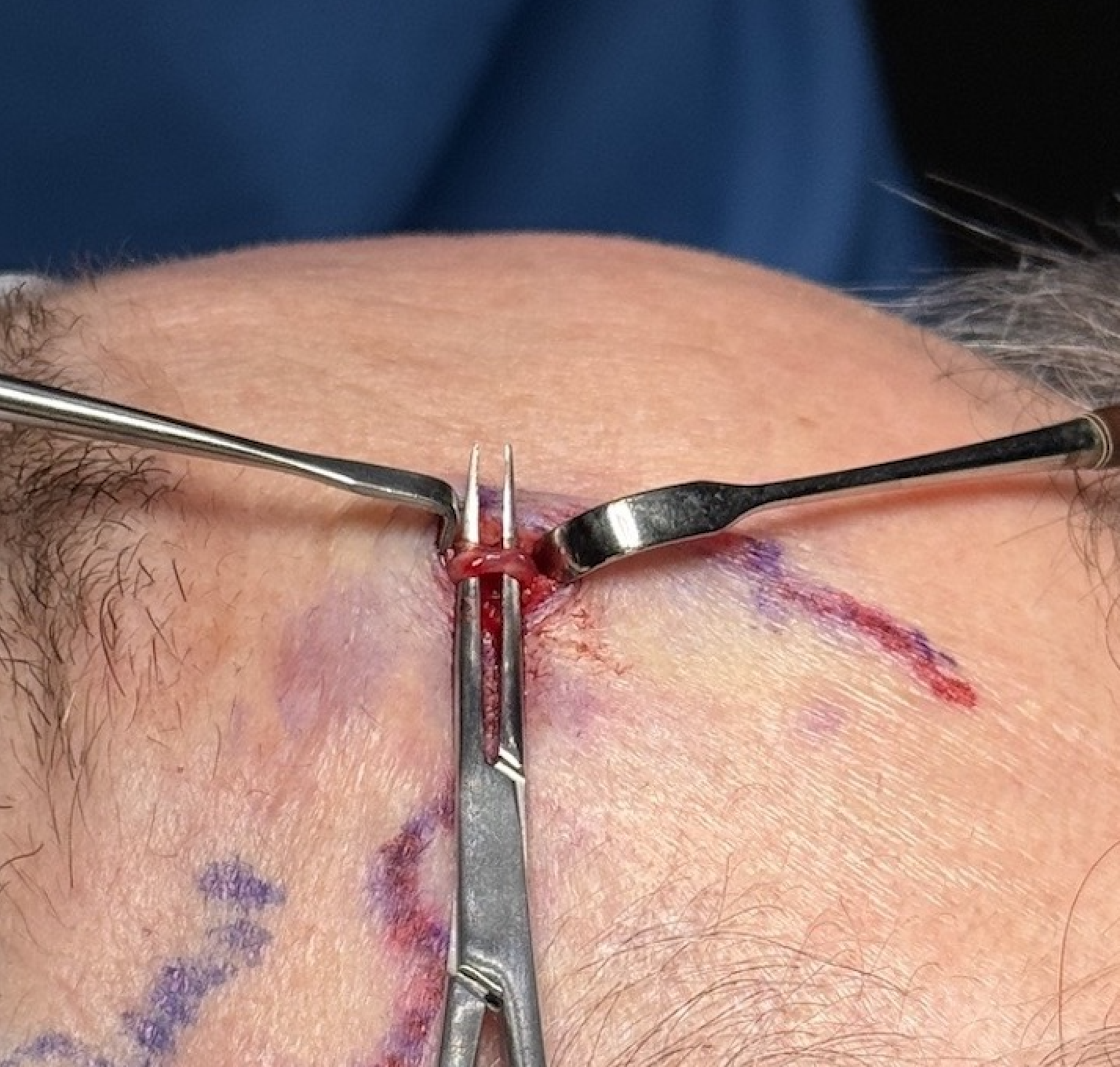
3. Identify the Artery Early
The STA is usually very superficial.
Technique:
- Small incision directly over the vessel
- Blunt spreading with tenotomy scissors
- Use NO electrocautery
The artery should appear before any deep fascia is encountered.
If you see white fascial fibers, you are already too deep.
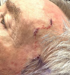
4. Incision Placement
Two approaches help protect the nerve:
Direct vessel incision
- 5 – 7mm incision directly over the arteryy
No matter where the incisions are placed it is safe if dissection stays subcutaneous.
5. Avoid High-Risk Areas
Greatest nerve vulnerability:
- 2–4 cm above the zygomatic arch
- Anterior temple
If ligating a frontal branch of the STA, be particularly careful in this region.
6. Blunt Dissection Only
Avoid sharp deep dissection.
Best tools:
- Mosquito clamp
- Tenotomy scissors
- Gentle spreading
Once the artery is isolated and pulled out (extracted) from the incision, then double ligate. Cutting between the suture ligations is an option but I usually don’t do it.
7. Visual Clues You Are Safe
You should see:
- Yellow fat
- Thin red artery
- No fascia or muscle fibers
If fascia appears, stop and come superficial again.
But extracting the artery from the incision, which can be done because of the natural slack that the superficial temporal artery has, offers assurance that all that is being ligated is the artery.
8. If the Nerve Is Injured
Clinical sign:
- Inability to raise the lateral eyebrow
- Forehead asymmetry
Most injuries in this area are neuropraxia and recover in 3–6 months, but prevention is obviously critical.
Key surgical pearl
The superficial temporal artery lies superficial to the nerve, so if you never leave the subcutaneous plane, the frontal branch of the facial nerve will remain protected.
Dr. Barry Eppley
Plastic Surgeon