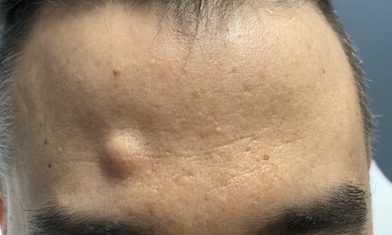


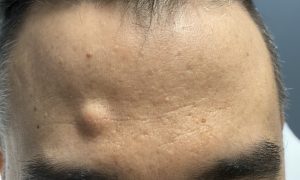
 Solid masses of the forehead appear as round immobile protrusions. They are visually unavoidable given the broad surface area of the forehead. They start out as small discrete round bumps and then gradually over time get bigger. How big they get is uncertain but anywhere from a the size of an eraser to a quarter in diameter is typical. While most can be directly removed through a small overlying skin incision a scar on the forehead is usually not appealing to most patients, particularly when there are no significant horizomtal wrinkle lines in which to place it.
In the October 2022 issue of the journal European Journal of Plastic Surgery an article on this topic was published entitled ‘Single Port Endoscopic Approach for Forehead Lesions: A Single-Center Case Series.’ In this paper the authors review their three year experience treating benign forehead masses with a single port (single scalp incision) endoscopic frontal resection technique. Sixteen (16) patients were treated with the removal of twenty (20) masses. The histology of the masses were lipomas (10), osteomas (8) and cysts. (2) Two complications occurred , a hematoma and a cyst recurrence.
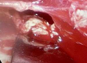
 While the endoscopic technique is used traditionally with two ports (a visualization port and a working port) and two separate incisions, I have never found that necessary when it comes to forehead mass removals. A single port allows both the endoscope and any instrument to be passed through the same small scalp incision to dissect out and remove the mass. Osteomas are sharply removed with an osteotome. Lipomas and cysts have to be bluntly dissected out and then grasped and removed.
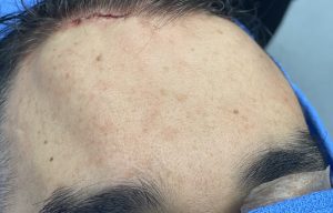
Such an endoscopic forehead removal technique, besides avoiding a forehead scar, does not take any more time to do than a direct overlying skin incision method.
Dr. Barry Eppley
World-Renowned Plastic Surgeon