

The S-shape of the clavicle bone is anchored by very strong ligamentous attachments at both ends to joints of the sternum and scapula. Because of its horizontal orientation it also serves as a location for multiple muscle attachments of the neck, chest and shoulder. These muscles include from medial to lateral the sternocleidomastoid, pectoralis major, subclavius, deltoid and trapezius.

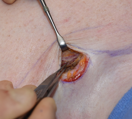
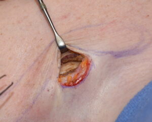 In performing shoulder narrowing surgery by shortening the length of the clavicle, surgical access is done through an incision at its medial third. To gain adequate subperiosteal exposure to the length of inner clavicle bone needed to perform both the bone removal and plate fixation, the pectoralis major and sternocleidomastoid muscle attachments will be encountered.
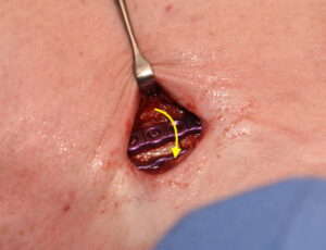
 Releasing enough of the pectoralis major and sternocleidomastoid muscles from the bone is necessary to complete the procedure. During closure the detached muscles are brought back over the bone and fixation plate and reapproximated together. This is done primarily to provide good soft tissue coverage/camouflage of the superior plate and screws.
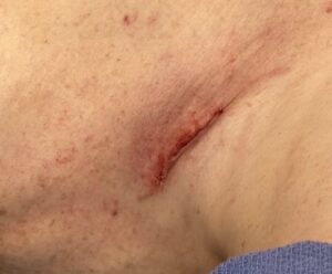
The one side effect of this muscle manipulation is that is actually the greatest source of postoperative discomfort. It is not the cutting of the bone but the detachment/reattachment of the pectoralis major muscle in particular that causes some persistent postoperative soreness. While this represents a relatively small area of pectoralis major muscle origin, which is much more extensive down along the sternum, it is an area that gets pulled on by elevation of the arm. This persists most significantly in the first few weeks after surgery but is largely resolved by four to six weeks when arm motion has largely returned to normal.
One of the interesting, but functionally insignificant, effects of bringing the released pectoralis major muscle section up over the fixation plate is that on flexion of the neck a band can be seen up along the course of the sternocleidomastoid muscle in some patients. This is the result of actually making a connection between the pectoralis and sternocleidomastoid muscle which are normally separated by the bone to which they are attached.
Dr. Barry Eppley
Indianapolis, Indiana