

Shoulder reduction is now a common procedure for those seeking a less wide or square shaped shoulders. This is done by removing a section of the straight portion of the clavicle and then reuniting the bones after the removal with plate and screw fixation. Since the clavicle a longitudinal bone that keeps the shoulder at a distance from the sternum it is no surprise that shortening its length changes the outer shape of the shoulders in a reductive manner. Such changes are seen immediately after the surgery as the swelling from the bone removal area is at a distance from the outer edge of the shoulders.
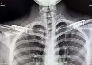
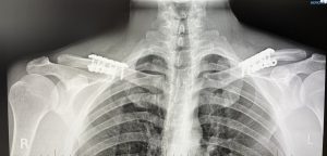
A postoperative x-ray is always taken within a day or two after the surgery for multiple reasons. One obvious reason is for documentation of the bone fix station site to verify its an alignment. While this is true that is not the most significant reason given that there is clear visibility of how two bone ends were put together with the plates and screws at the time of surgery. It would have been very unlikely that any changes would have occurred in the osteotomy fixation site within a day or two after the surgery unless a significant traumatic event had occurred… which would be well known to the patient.
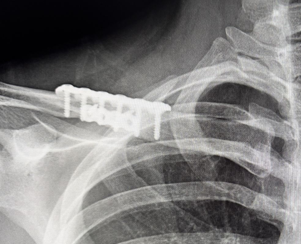
 My primary motivation and giving a postoperative x-ray is to check the length of the screws and to verify that good bicortical engagement of them had occurred. In surgery the actual length of the screws needed to ensure that they go through both sides of the bony cortical layers is an estimate based on measurements of the bone that was removed. The primary reason fixation n of the clavicle fails in reduction osteotomies is, which is very rare in my experience, is due to screw pullout. This is far difficult to occur when the threads of the screw engage both the superior and inferior cortical bone layers adequately. It is easy to check on the x-ray whether the end of the screws goes slightly past the inferior cortical layer on the bottom of the bone which is desired. Such screw tip location cannot be seen for most of the screws doing surgery.
 Besides the very visible hardware there are other interesting findings that can be seen on postoperative x-rays. Perhaps not unsurprisingly there is an increased space in the acromioclavicular joint which u doubtably is due to the inward pull of the lateral clavicle bone segment as the shoulders are narrowed. It is unclear weather this increase joint space persist but I would suspect that this is a short term change and the joint space over time returns to its normal size.
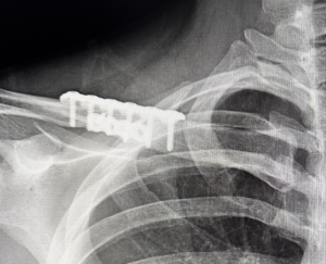 Also, although more difficult to see, is the discrepancy in the size of the two bones segments that have been put back together. The outer clavicle bone segment is usually only about 2/3 the size of the inner bone segment to which it is aligned. It is amazing how much the clavicle narrows as it extends out laterally.While these diameter differences do not affect how the bone heals one does wonder long term if the actual strength of the clavicle at the osteotomy site ever assumes what it was with an equal bone diameter before the surgery.
The postoperative x-ray is a valuable tool after clavicle reduction surgery to both assess the hardware used to do it as well as observe other interesting bone and joint changes.
Dr. Barry Eppley
World-Renowned Plastic Surgeon