


Background
Among aesthetic muscle augmentation procedures, trapezius implants are among the least performed. Their rarity is underscored by the absence of commercially available trapezius implants, indicating low patient demand. Surgeons seeking to augment this muscle typically rely on implants designed for other areas, such as calf implants, or commission custom implants tailored to each patient. Beyond the limited implant options, the procedure itself is so uncommon that no standardized surgical technique exists.
The trapezius is a large back muscle divided into three sections according to origin and insertion points:
- Upper trapezius: Extends from the base of the skull to the shoulder joint (acromion process and clavicle).
- Middle trapezius: Originates from the lower cervical and upper thoracic vertebrae, inserting onto the scapular spine.
- Lower trapezius: Originates from the lower thoracic vertebrae, also inserting onto the scapular spine.
Of these, only the upper trapezius holds aesthetic significance, as it contributes to both frontal and posterior views and complements the deltoid muscle in achieving a balanced, muscular appearance.
Surgical Approaches
Two primary approaches exist for trapezius implant placement:
- Medial high-to-low approach – involving an incision at the base of the neck.
- Lateral high-to-low approach – involving an incision near the acromio-clavicular (AC) joint.
These approaches differ in incision placement, intraoperative positioning, and implant orientation. The following case illustrates both methods, performed sequentially on the same patient.
Case Study
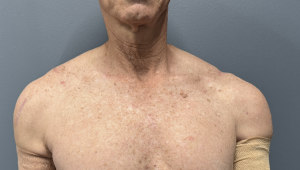
An older male patient, who had previously undergone pectoral, bicep, tricep, and deltoid augmentations, sought to complete his upper body enhancement with upper trapezius augmentation.
First Procedure (Medial Approach):
 Performed in the prone position.
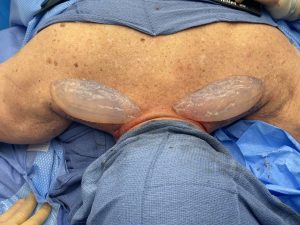
- A low medial incision was made, and a pocket was developed over the muscle fascia extending to the AC joint.
- Modified calf implants, fuller near the neck and tapering toward the shoulder, were inserted.
- The patient healed uneventfully, with improved definition of the upper trapezius in both frontal and posterior views.
 Performed in the prone position.
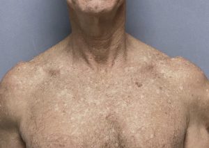
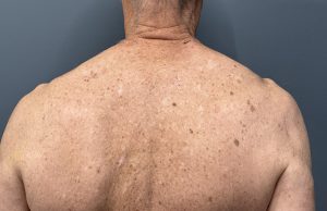 The patient healed uneventfully, with improved definition of the upper trapezius in both frontal and posterior views.
Second Procedure (Lateral Approach):
- One year later, the patient requested a more natural contour, with reduced fullness at the neck and increased volume near the shoulder.
- Using a lateral incision at the AC joint, the original implants were removed.
- New modified calf implants were placed, designed to be thicker near the incision and thinner toward the neck.
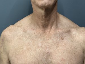
- Postoperatively, the patient expressed a preference for the new trapezius profile with a lower neck profile and more fullness that extended out closer to the shoulder.
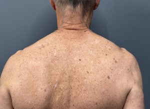
 Postoperatively, the patient expressed a preference for the new trapezius profile with a lower neck profile and more fullness that extended out closer to the shoulder.
Outcomes and Considerations
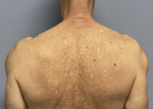
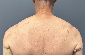
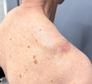 Comparison of the two procedures revealed distinct differences in contour. The second set of implants produced a more natural and aesthetically pleasing result according to the patient. Healing of the lateral incision was favorable, with minimal risk of becoming an aesthetic concern. In fact, lateral incisions can be attributed to orthopedic shoulder procedures, allowing patients discretion if they prefer not to disclose cosmetic surgery.
Key Insights
- Trapezius implants may be placed via either a medial low-neck incision or a lateral AC joint incision.
- Implant design can emphasize fullness near the neck or the shoulder joint, depending on patient preference.
- While custom implants can be fabricated, modified calf implants remain a practical and effective alternative.
Dr. Barry Eppley
World-Renowned Plastic Surgeon