

Cheekbone reduction surgery requires an understanding of the complete bony anatomy of the zygomatic bone. When most people think of the cheekbone ti is perceived as one solid block of bone just underneath the eye. While this area is a major part of the cheekbone, it overlooks the posterior extension of the cheekbone known as the zygomatic arch.
The zygomatic arch connects the main body of the cheekbone (zygoma) to the temporal bone above the ear. It is a thin bridge of bone between these two areas because underneath it passes the large temporalis muscle on its ways to attach to the lower jaw. The zygomatic arch is almost always bowed outward or has a convex shape. This gives width to the side of the midface.
In cheekbone reduction it is rarely a matter of shaving down the bone. Rather the cheekbone is cut and moved inward, this is what make the side of the face more narrow. The bone cuts are done in the front through the main body of the zygoma from an intraoral incision. Conversely the back cut is done where the zygomatic arch meets the temporal bone through an external incision.
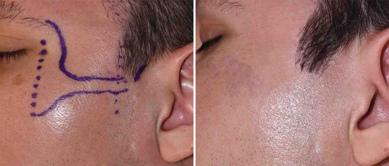 The external incision for the zygomatic arch osteotomy is done through the sideburn hair. It is usually about 1 cm in length and is placed at the junction of the sideburn hair and skin just in front of the ear. Because it is an external incision patients understandably are concerned about how it heals an whether it heals in an inconspicuous manner. Here is a picture of a patient with a posterior zygomatic arch osteotomy incision that was done just over one year ago.
Dr. Barry Eppley
Indianapolis, Indiana