The prevalence of migraine headaches, specifically those that are disabling and life-altering, is not rare. When traditional neurologic approaches, such as drugs destined to prevent or abort a migraine, fail to provide relief through a centrally-mediated mechanism then peripheral therapy should be considered. A lot of new information is forthcoming that supports the peripheral theory of certain migraines caused by external trigger points that then migrate centrally to the brain. This has spawned trigger point therapy using an injectable drug or surgery.Through either Botox injections or decompression of the brow (supraorbital) or base of the skull (occipital) nerves as they exit from their muscular beds, significant and sustained relief has been obtained in selected migraine sufferers.
But not every migraine responds to these peripheral muscular decompression treatments norc an certain migraines even be explained by such approaches. One specific type of migraine emanates from pain high in the temple (temporal) region. The image of someone holding their temples while in pain is a classic one for migraines. Some patients will tell you that they can make it actually feel better, experience less pain, if they press in on their temple area. Whether this maneuver works is unknown but some patients feel that it helps. Migraines in the temple region do not appear to have a muscular trigger point as there is no specific cranial nerve in the temple area that passes out through the muscle like that in the brow or at the back of the heads.
One potential explanation is that the trigger in the temporal area may be medicated by a vascular origin and not muscle. The auriculotemporal nerve passes through the temple area, largely being on top of the muscle. Its pathway is located fairly close to where most temporal migraine sufferers can put their finger on as the most intense areas of pain. This type of pain location is where both the auriculotemporal nerve) and the superficial temporal artery are in close association.
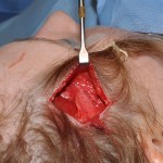
Recent published anatomic studies by plastic surgeons has shown that up to one-third of cadaver head dissected showed a direct relationship between the artery and the nerve where they crossed each other or became actually intertwined. This could be a source of nerve irritation and a potential trigger for temporal-based headaches. As the nerve and artery are located in the superficial fascia of the temple, they both are easily accessible through very limited incisions inside the temporal hairline.

The field of migraine surgery through muscle or vascular decompression is new and continues to evolve. Less studies have been done on temporal migraine decompression but vascular ligation/separation seems like a plausible theory. There are virtually no downsides to this procedure other than it may not work. It is simple enough that it can be done under local anesthesia if it is the only trigger point being decompressed. Diagnostic testing of the temple region as a potential culprit can not be done by Botox to prove the vascular trigger point theory or that the surgery may be effective. Local anesthetic injection would seem to more reliable as a nerve block test but one has to be careful to not puncture the underlying artery. The correlation between a positive local anesthetic nerve block and the success of vascular decompression, however, has not been proven and further studies are needed.
Dr. Barry Eppley
Indianapolis, Indiana


