

Widening of the side of the head is possible through the use of specially designed implants. Known as posterior temporal implants, they specifically augment the convex portion of the temporal bone above the ears.
Understanding the anatomy of the temporal bone allows one to understand how this is possible. The temporal bone has four distinct areas which vary greatly in their shape. The squamosal portion of the bone is its largest and most superiorly positioned part sitting above the ears. This is also its most convex part. The smaller zygomatic part is the anterior projection that sticks forward to reach to the zygomatic bone. The mastoid and petrous parts extend below the squamous part and are irrelevant for this discussion.
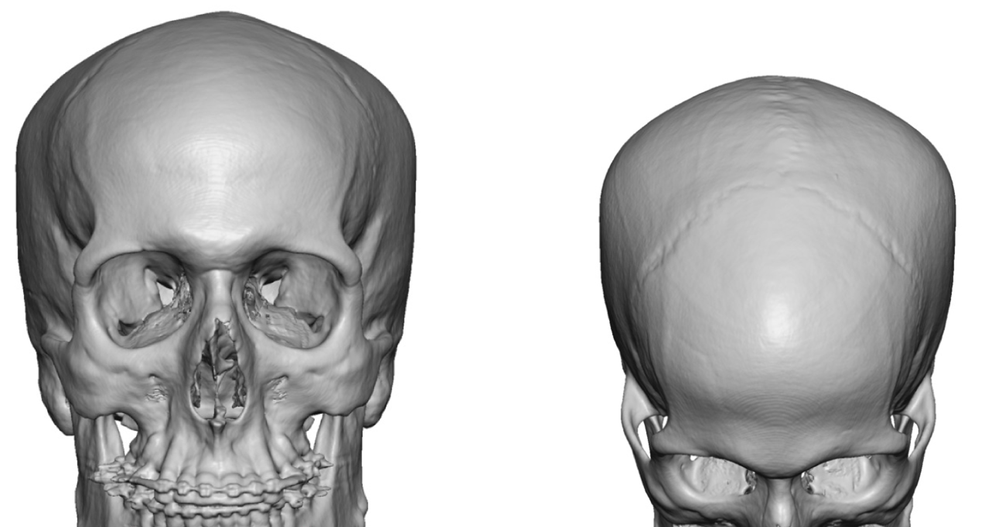 It can be seen that what I refer to as the posterior temporal area, as differentiated by anterior temporal augmentation by the side of the eye, is the squamosal part of the temporal bone. The normal convex shape of the bone is usually flat in individuals that desire head widening as seen in the attached 3D imaging.
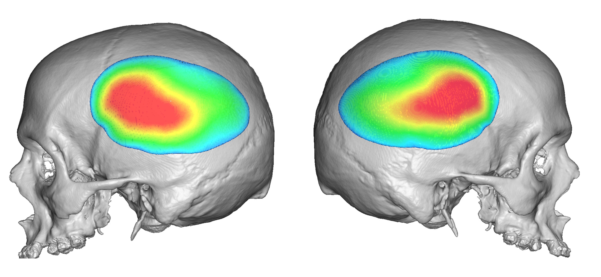 The design of the head widening implant is oblong in shape. It is designed to provide augmentation well above the external auditory canal. Its maximum projection should be at or just above the level of the eye. It has a feathering design from the central projected area to allow for a smooth shape into the surrounding areas. Its typical augmentation amount is 5mm to 7mms although much larger amounts can and has been done.
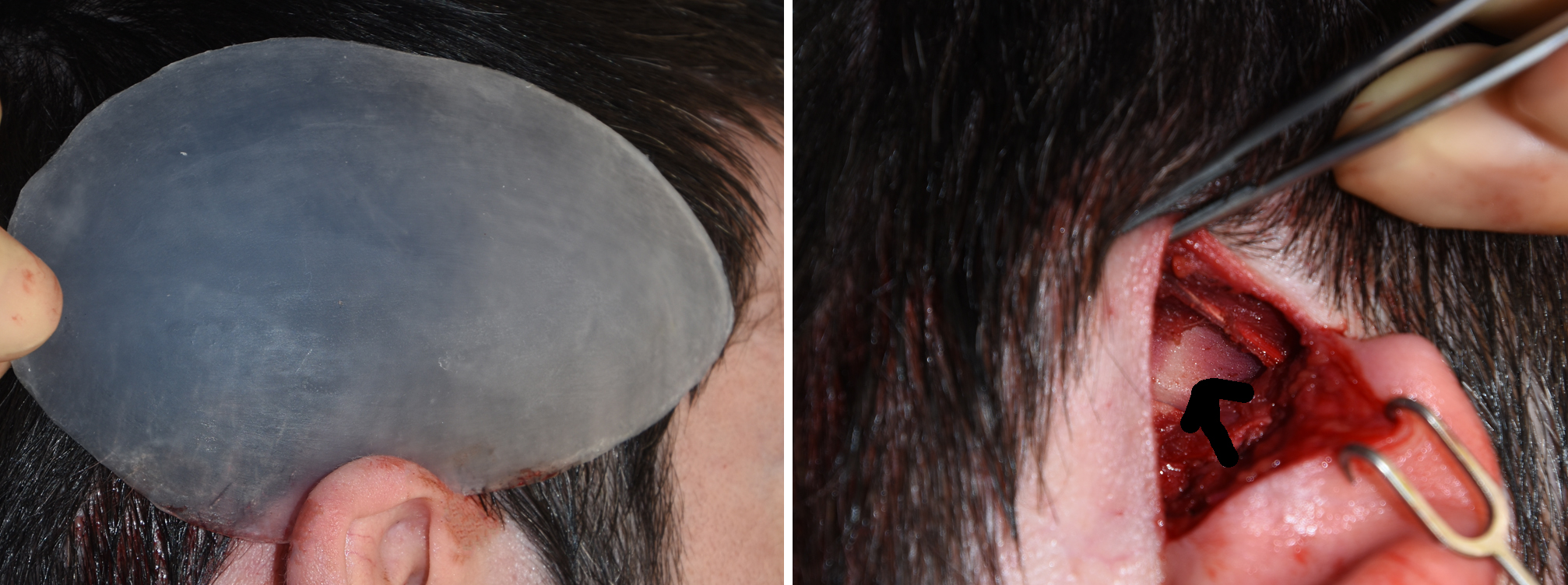 The head widening implant is placed through a postauricular incision in a submuscular pocket. This is the most effective approach to pushing out the side of the head with the posterior belly of the temporal muscle on top of the implant. It is secured with a single screw to keep it in its desired position.
Dr. Barry Eppley
Indianapolis, Indiana